Ankle
Editor-In-Chief: C. Michael Gibson, M.S., M.D. [1]
In human anatomy, the ankle joint is formed where the foot and the leg meet. The ankle, or talocrural joint, is a synovial hinge joint that connects the distal ends of the tibia and fibula in the lower limb with the proximal end of the talus bone in the foot.[1] The articulation between the tibia and the talus bears more weight than between the smaller fibula and the talus.
The term "ankle" is used to describe structures in the region of the ankle joint proper.[2]
Movement
The ankle joint is responsible for dorsiflexion (moving the toes up as when standing only on the heels) and plantar flexion of the foot (moving the toes down, as when standing on the toes), and allows for the greatest movement of all the joints in the foot. The ankle does not allow rotation.
In plantar flexion, the anterior ligaments of the joint become longer while the posterior ligaments become shorter. The reverse is true for dorsiflexion.
Articulation
The lateral malleolus of the fibula and the medial malleolus of the tibia along with the inferior surface of the distal tibia articulate with three facets of the talus. These surfaces are covered by cartilage.
The anterior talus is wider than the posterior talus. When the foot is dorsiflexed , the wider part of the superior talus moves into the articulating surfaces of the tibia and fibula, creating a more stable joint than when the foot is plantar flexed.
Ligaments
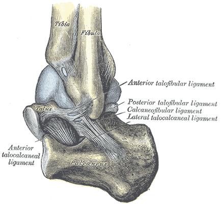
The ankle joint is bound by the strong deltoid ligament and three lateral ligaments: the anterior talofibular ligament, the posterior talofibular ligament, and the calcaneofibular ligament.
- The deltoid ligament supports the medial side of the joint, and is attached at the medial malleolus of the tibia and connect in four places to the sustentaculum tali of the calcaneus, calcaneonavicular ligament, the navicular tuberosity, and to the medial surface of the talus.
- The anterior and posterior talofibular ligaments support the lateral side of the joint from the lateral malleolus of the fibula to the dorsal and ventral ends of the talus.
- The calcaneofibular ligament is attached at the lateral malleolus and to the lateral surface of the calcaneus.
The joint is most stable in dorsiflexion and a sprained ankle is more likely to occur when the foot is plantar flexed. This type of injury more frequently occurs at the anterior talofibular ligament.
Name derivation
The word ankle or ancle is common, in various forms, to Germanic languages, probably connected in origin with the Latin "angulus", or Greek "αγκυλος", meaning bent.
Related terms
A common variant of the word "ankle" is "cankle", which is commonly used derogatorily to describe the ankles of obese individuals where the ankle and calf may be indistinguishable. This insult can be taken further (often jokingly) using the term "thankle", which implies a person's thighs and ankles are indistinguishable.
Fractures

Most traumatic incidents involving the ankle result in ankle sprains. Symptoms of an ankle fracture can be similar than for sprains (pain, hematoma) or there may be an abnormal position, abnormal movement or lack of movement (if there is an accompanying dislocation), or the patient may have heard a crack.
On clinical examination, it is important to evaluate the exact location of the pain, the range of motion and the condition of the nerves and vessels. It is important to palpate the calf bone (fibula) because there may be an associated fracture, and to palpate the sole of the foot to look for a Jones fracture.
Evaluation of ankle injuries for fracture is done with the Ottawa ankle rules, a set of rules that were developed to minimize unnecessary X-rays. On X-rays, there can be a fracture of the medial malleolus, the lateral malleolus, or the anterior or posterior margin. If both malleoli are broken, this is called a bimalleolar fracture (some of them are called Pott's fractures). If three of these are broken at the same time, this is called a trimalleolar fracture (although there are only two malleoli). Ankle fractures are classified according to Weber, depending on their position relative to the anterior ligament of the lateral malleolus (type A = below the ligament, type B = at its level, type C = above the ligament). A special form of type C fracture is the Maisonneuve fracture, which involves a spiral fracture of the fibula with a tear of the distal tibiofibular syndesmosis and the interosseous membrane.
Only type A fractures of the lateral malleolus can be treated like sprains; all other types require surgery (most often an open reduction and internal fixation). A cast may be required to immobilize the ankle following surgery. Trimalleolar fractures or those with dislocation have a high risk of developing arthrosis.
Additional images
-
The bones in the foot.
-
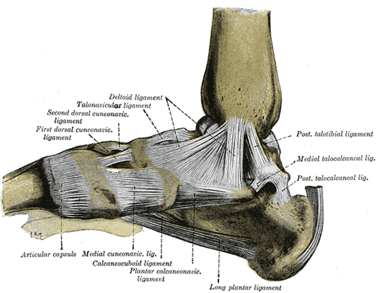 Ligaments of the medial aspect of the foot.
 -
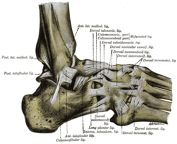 The ligaments of the foot from the lateral aspect.
 -
 Capsule of left talocrura articulation (distended). Lateral aspect.
 -
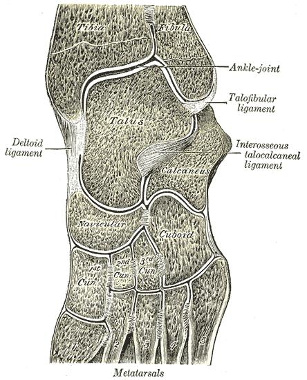 Oblique section of left intertarsal and tarsometatarsal articulations, showing the synovial cavities.


References
- ↑ "eMedicine/Stedman Medical Dictionary Lookup!". Retrieved 2008-01-14.
- ↑ "eMedicine/Stedman Medical Dictionary Lookup!". Retrieved 2008-01-14.
- Calais-Germain, Blandine. "Anatomy of Movement", Eastland Press, 1993. ISBN 0-939616-17-3
- Martini, Frederic; Timmons, Michael; McKinnley, Michael. "Human Anatomy", 3rd Edition, Prentice-Hall, 2000. ISBN 0-13-010011-0
- Marieb, Elaine. "Essentials of Human Anatomy and Physiology", 6th Edition. Addison Wesley Longman, 2000. ISBN 0-8053-4940-5
See also
| File:Wiktionary-logo-en-v2.svg | Look up ankle in Wiktionary, the free dictionary. |
External links
| Wikimedia Commons has media related to Ankle. |
- The Ankle from the University of Glasgow
- How to Diagnose Lateral Ankle Injuries from Podiatry Today
- American Academy of Orthopedic Surgeons website about Foot & Ankle
Template:Joints of lower limbs Template:Human anatomical features