Hodgkin's lymphoma pathophysiology
|
Hodgkin's lymphoma Microchapters |
|
Diagnosis |
|---|
|
Treatment |
|
Case Studies |
|
Hodgkin's lymphoma pathophysiology On the Web |
|
American Roentgen Ray Society Images of Hodgkin's lymphoma pathophysiology |
|
Risk calculators and risk factors for Hodgkin's lymphoma pathophysiology |
Editor-In-Chief: C. Michael Gibson, M.S., M.D. [1]; Associate Editor(s)-in-Chief: Sowminya Arikapudi, M.B,B.S. [2]
Overview
On gross pathology, white-grey, uniform, and enlarged lymph nodes are characteristic findings of Hodgkin's lymphoma. On microscopic histopathological analysis, Reed-Sternberg cells, reactive cell infiltrate, and complete or partial effacement of the lymph node architecture are characteristic findings of Hodgkin's lymphoma.
Pathophysiology
Hodgkin's lymphoma, is a type of lymphoma, in which cancer originates from a specific type of white blood cells called lymphocytes.[1][2]
Associated Conditions
Reports from countries like Honduras,[3] China,[4] Mexico,[5] Peru,[6] and Malaysia[7] suggest an association between EBV infection and Hodgkin's lymphoma, an association that is more evident in the pediatric population[8] and in the subtype of mixed cellularity.[9]
Gross Pathology
On gross pathology, affected lymph nodes (most often, latero cervical lymph nodes) are enlarged, but their shape is preserved because the capsule is not invaded. Usually, the cut surface is white-grey and uniform. In some histological subtypes (e.g. nodular sclerosis), the cut surface may have a nodular aspect.
-
![Hodgkin lymphoma - Massive involvement of para tracheal, hilar and sub carinal lymph nodes as well as two vertebral bodies.[10]](/images/8/8a/Hodgkin%27s_lymphoma_Gross_Pathology.jpg)
Hodgkin lymphoma - Massive involvement of para tracheal, hilar and sub carinal lymph nodes as well as two vertebral bodies.[10]
![Hodgkin lymphoma - Massive involvement of para tracheal, hilar and sub carinal lymph nodes as well as two vertebral bodies.[10]](/index.php/File:Hodgkin%27s_lymphoma_Gross_Pathology.jpg)
Microscopic Pathology
Microscopic examination of the lymph node biopsy reveals complete or partial effacement of the lymph node architecture by scattered large malignant cells known as Reed-Sternberg cells (typical and variants) admixed within a reactive cell infiltrate composed of variable proportions of lymphocytes, histiocytes, eosinophils, and plasma cells. The Reed-Sternberg cells are identified as large often bi-nucleated cells with prominent nucleoli and an unusual CD45-, CD30+, and CD15+/- immuno phenotype. In approximately 50% of cases, the Reed-Sternberg cells are infected by the Epstein-Barr virus.
-
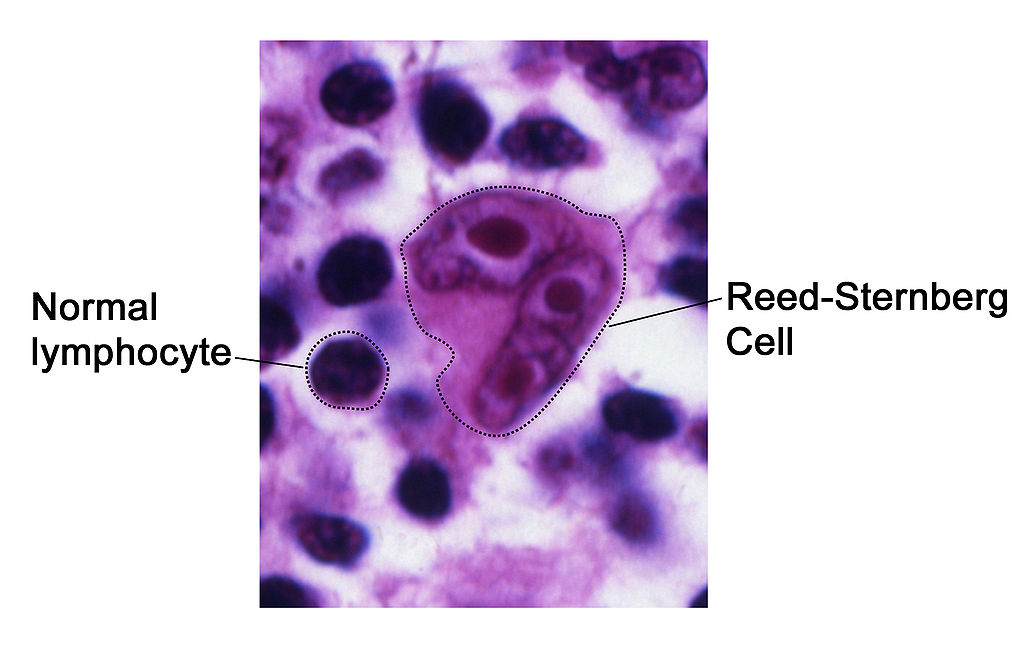
Reed-Sternberg Hodgkin's Lymphoma
-
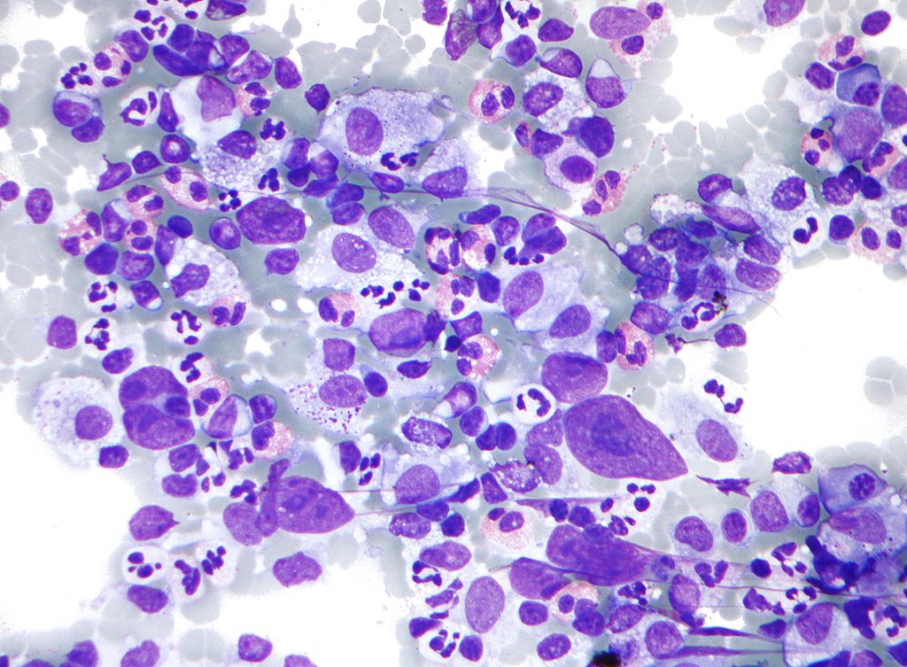
Micrograph of Hodgkin lymphoma, abbreviated HL. Lymph node FNA specimen. Field stain. The micrograph shows a mixture of cells common in Hodgkin's lymphoma: eosinophils, Reed-Sternberg cells, plasma cells, and histocytes.
-
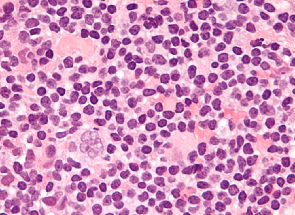
Micrograph showing a "popcorn cell", the Reed–Sternberg cell variant seen in nodular lymphocyte predominant Hodgkin lymphoma. H&E stain


| Type of cell | Characteristics | |
|---|---|---|
| Classic | ||
| Reed-Sternberg cells (RSC) | Include large size (20–50 micro metres), abundant, amphophilic, finely granular/homogeneous cytoplasm; two mirror-image nuclei (owl eyes) each with an eosinophilic nucleolus and a thick nuclear membrane (chromatin is distributed close to the nuclear membrane). | |
| Variants | ||
| Hodgkin cells (Atypical mononuclear Reed-Sternberg cell) | Have the same characteristics as Reed-Sternberg cells (RSC), but is mono nucleated. | |
| Lacunar Reed-Sternberg cells | Have a single hyper lobulated nucleus, multiple, small nucleoli and eosinophilic cytoplasm which is retracted around the nucleus, creating an empty space ("lacunae"). | |
| Pleomorphic Reed-Sternberg cells | Have multiple irregular nuclei. | |
| "Popcorn" Reed-Sternberg cells (Lympho-histiocytic variant) | Have a very lobulated nucleus and small nucleoli. | |
| "Mummy" Reed-Sternberg cells | Have a compact nucleus with no nucleolus and basophilic cytoplasm. | |
References
- ↑ Scientific Style and Format: The CBE Manual for Authors, Editors, and Publishers. Cambridge University Press. 1994. pp. 97–. ISBN 978-0-521-47154-1.
- ↑ Lozano R, Naghavi M, Foreman K, Lim S, Shibuya K, Aboyans V, et al. (Dec 15, 2012). "Global and regional mortality from 235 causes of death for 20 age groups in 1990 and 2010: a systematic analysis for the Global Burden of Disease Study 2010". Lancet. 380 (9859): 2095–128. doi:10.1016/S0140-6736(12)61728-0. OCLC 23245604.
- ↑ Ambinder RF, Browning PJ, Lorenzana I, Leventhal BG, Cosenza H, Mann RB; et al. (1993). "Epstein-Barr virus and childhood Hodgkin's disease in Honduras and the United States". Blood. 81 (2): 462–7. PMID 8380725.
- ↑ Zhou XG, Hamilton-Dutoit SJ, Yan QH, Pallesen G (1993). "The association between Epstein-Barr virus and Chinese Hodgkin's disease". Int J Cancer. 55 (3): 359–63. PMID 8397160.
- ↑ Zarate-Osorno A, Roman LN, Kingma DW, Meneses-Garcia A, Jaffe ES (1995). "Hodgkin's disease in Mexico. Prevalence of Epstein-Barr virus sequences and correlations with histologic subtype". Cancer. 75 (6): 1360–6. PMID 7882287.
- ↑ Chang KL, Albújar PF, Chen YY, Johnson RM, Weiss LM (1993). "High prevalence of Epstein-Barr virus in the Reed-Sternberg cells of Hodgkin's disease occurring in Peru". Blood. 81 (2): 496–501. PMID 8380728.
- ↑ Peh SC, Looi LM, Pallesen G (1997). "Epstein-Barr virus (EBV) and Hodgkin's disease in a multi-ethnic population in Malaysia". Histopathology. 30 (3): 227–33. PMID 9088951.
- ↑ Armstrong AA, Alexander FE, Paes RP, Morad NA, Gallagher A, Krajewski AS; et al. (1993). "Association of Epstein-Barr virus with pediatric Hodgkin's disease". Am J Pathol. 142 (6): 1683–8. PMC 1886981. PMID 8389527.
- ↑ Andriko JA, Aguilera NS, Nandedkar MA, Abbondanzo SL (1997). "Childhood Hodgkin's disease in the United States: an analysis of histologic subtypes and association with Epstein-Barr virus". Mod Pathol. 10 (4): 366–71. PMID 9110300.
- ↑ Image courtesy of Dr Yale Rosen Radiopaedia(original file ‘’here’’). Creative Commons BY-SA-NC