HIV AIDS chest x ray: Difference between revisions
Jump to navigation
Jump to search
m (Bot: Removing from Primary care) |
|||
| (20 intermediate revisions by 5 users not shown) | |||
| Line 1: | Line 1: | ||
__NOTOC__ | __NOTOC__ | ||
{{AIDS}} | {{AIDS}} | ||
{{CMG}}; '''Associate | {{CMG}}; '''Associate Editor-in-Chief:'''{{SSK}} | ||
==Overview== | ==Overview== | ||
Chest X-ray | Chest X-ray findings in HIV/AIDS are related to the development of opportunistic lung infections. They include ground-glass infiltrates suggestive of ''Pneumocystis jirovecii'' pneumonia, lobar consolidation, pleural effusions, loculated empyemas, and lymphadenopathy. | ||
==Chest X Ray== | ==Chest X Ray Findings== | ||
Chest X-ray findings in HIV/AIDS are related to the development of opportunistic lung infections. Common findings include:<ref name="pmid20981180">{{cite journal| author=Allen CM, Al-Jahdali HH, Irion KL, Al Ghanem S, Gouda A, Khan AN| title=Imaging lung manifestations of HIV/AIDS. | journal=Ann Thorac Med | year= 2010 | volume= 5 | issue= 4 | pages= 201-16 | pmid=20981180 | doi=10.4103/1817-1737.69106 | pmc=PMC2954374 | url=http://www.ncbi.nlm.nih.gov/entrez/eutils/elink.fcgi?dbfrom=pubmed&tool=sumsearch.org/cite&retmode=ref&cmd=prlinks&id=20981180 }} </ref> | |||
*'''Diffuse ground-glass infiltrates''' | |||
:* Suggestive of ''Pneumocystis jirovecii'' pneumonia | |||
*'''Nodular infiltrates''' | |||
:* Suggestive of bacterial or fungal pneumonia | |||
*'''Lobar/segmental consolidation''' | |||
:* Suggestive of bacterial or fungal pneumonia | |||
*'''Pleural effusion''' | |||
:* Suggestive of empyema, parapneumonic effusion, tuberculous effusion, and malignant effusion | |||
*'''Lobar consolidation''' | |||
:* Suggestive of bacterial or fungal pneumonia | |||
*'''Hilar lymphadenopathy''' | |||
:* Suggestive of tuberculosis, malignancy, or may be secondary to HIV induced lymphadenopathy | |||
*'''Cavitation''' | |||
:* Suggestive of tuberculosis, fungal infection, or necrotizing pneumonia | |||
*'''Mass lesion''' | |||
:* Suggestive of malignancy, tuberculosis, or fungal infection | |||
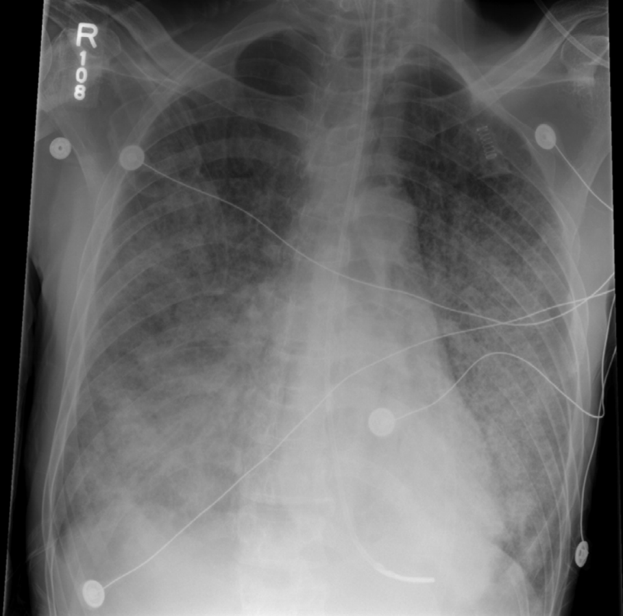
[[File:Pneumocystis_jirovecii_pneumonia_CXR.png|thumb|none|500px|Chest X-ray of an individual with ''Pneumocystis jirovecii'' pneumonia<ref name="pmid22096390">{{cite journal| author=Castro JG, Morrison-Bryant M| title=Management of Pneumocystis Jirovecii pneumonia in HIV infected patients: current options, challenges and future directions. | journal=HIV AIDS (Auckl) | year= 2010 | volume= 2 | issue= | pages= 123-34 | pmid=22096390 | doi= | pmc=PMC3218692 | url=http://www.ncbi.nlm.nih.gov/entrez/eutils/elink.fcgi?dbfrom=pubmed&tool=sumsearch.org/cite&retmode=ref&cmd=prlinks&id=22096390 }} </ref>]] | |||
=== | |||
==References== | ==References== | ||
{{reflist|2}} | {{reflist|2}} | ||
{{WH}} | |||
{{WS}} | |||
[[Category:HIV/AIDS]] | [[Category:HIV/AIDS]] | ||
[[Category:Disease]] | [[Category:Disease]] | ||
[[Category:Immune system disorders]] | [[Category:Immune system disorders]] | ||
[[Category: | [[Category:Viral diseases]] | ||
[[Category:Pandemics]] | [[Category:Pandemics]] | ||
[[Category:Sexually transmitted infections]] | [[Category:Sexually transmitted infections]] | ||
| Line 33: | Line 43: | ||
[[Category:Immunodeficiency]] | [[Category:Immunodeficiency]] | ||
[[Category:Microbiology]] | [[Category:Microbiology]] | ||
[[Category:Emergency mdicine]] | |||
[[Category:Up-To-Date]] | |||
[[Category:Infectious disease]] | |||
Latest revision as of 22:11, 29 July 2020
|
AIDS Microchapters |
|
Diagnosis |
|
Treatment |
|
Case Studies |
|
HIV AIDS chest x ray On the Web |
|
American Roentgen Ray Society Images of HIV AIDS chest x ray |
Editor-In-Chief: C. Michael Gibson, M.S., M.D. [1]; Associate Editor-in-Chief:Serge Korjian M.D.
Overview
Chest X-ray findings in HIV/AIDS are related to the development of opportunistic lung infections. They include ground-glass infiltrates suggestive of Pneumocystis jirovecii pneumonia, lobar consolidation, pleural effusions, loculated empyemas, and lymphadenopathy.
Chest X Ray Findings
Chest X-ray findings in HIV/AIDS are related to the development of opportunistic lung infections. Common findings include:[1]
- Diffuse ground-glass infiltrates
- Suggestive of Pneumocystis jirovecii pneumonia
- Nodular infiltrates
- Suggestive of bacterial or fungal pneumonia
- Lobar/segmental consolidation
- Suggestive of bacterial or fungal pneumonia
- Pleural effusion
- Suggestive of empyema, parapneumonic effusion, tuberculous effusion, and malignant effusion
- Lobar consolidation
- Suggestive of bacterial or fungal pneumonia
- Hilar lymphadenopathy
- Suggestive of tuberculosis, malignancy, or may be secondary to HIV induced lymphadenopathy
- Cavitation
- Suggestive of tuberculosis, fungal infection, or necrotizing pneumonia
- Mass lesion
- Suggestive of malignancy, tuberculosis, or fungal infection

References
- ↑ Allen CM, Al-Jahdali HH, Irion KL, Al Ghanem S, Gouda A, Khan AN (2010). "Imaging lung manifestations of HIV/AIDS". Ann Thorac Med. 5 (4): 201–16. doi:10.4103/1817-1737.69106. PMC 2954374. PMID 20981180.
- ↑ Castro JG, Morrison-Bryant M (2010). "Management of Pneumocystis Jirovecii pneumonia in HIV infected patients: current options, challenges and future directions". HIV AIDS (Auckl). 2: 123–34. PMC 3218692. PMID 22096390.