Dislocated patella
|
Dislocated patella Microchapters |
|
Diagnosis |
|---|
|
Treatment |
|
Case Studies |
|
Dislocated patella On the Web |
|
American Roentgen Ray Society Images of Dislocated patella |
Editor-In-Chief: C. Michael Gibson, M.S., M.D. [1]; Associate Editor(s)-in-Chief: Grace M. Gibson
Synonyms and keywords: dislocated kneecap; patellar dislocation; patellar subluxation; subluxation of the patella; subluxation of patella; luxating patella
Overview
Luxating patella, or trick knee, is a condition in which the patella, or kneecap, dislocates or moves out of its normal location. The luxation is usually medial, but can be lateral.
Classification
There are four diagnostic grades of patellar luxation[1], each more severe than the previous:
- Grade I - the patella can be manually luxated but is reduced (returns to the normal position) when released;
- Grade II - the patella can be manually luxated or it can spontaneously luxate with flexion of the stifle joint. The patella remains luxated until it is manually reduced or when the animal extends the joint and derotates the tibia in the opposite direction of luxation;
- Grade III - the patella remains luxated most of the time but can be manually reduced with the stifle joint in extension. Flexion and extension of the stifle results in reluxation of the patella;
- Grade IV - the patella is permanently luxated and cannot be manually repositioned. There may be up to 90¼ of rotation of the proximal tibial plateau. The femoral trochlear groove is shallow or absent, and there is displacement of the quadriceps muscle group in the direction of luxation.
Pathophysiology
It can be caused by some form of blunt trauma, or may be a congenital defect. In congenital cases, it is usually bilateral.
Natural History, Complications and Prognosis
Osteoarthritis can develop secondarily.
Diagnosis
Symptoms
Symptoms can range from none to severe pain.
Physical Examination
Diagnosis is made through palpation of the knee.
X-Rays
X-rays are necessary in some cases.
MRI
-
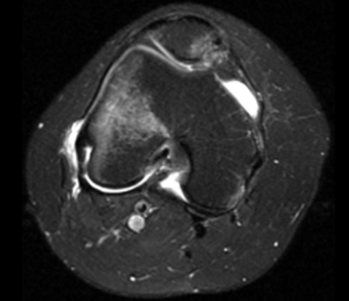
Lateral patellar dislocation Image courtesy of RadsWiki and copylefted

Treatment
Acute Care
Icing the joint can minimize swelling acutely.
Chronic Care
Grades III and IV, as well as most grade II cases, require surgery to correct, if the animal has difficulty walking. The surgery involves a sulcoplasty, a deepening of the trochlear sulcus that the patella sits in.
References
- ↑ OFA. "Patellar Luxation" (text/html). Orthopedic Foundation for Animals. Retrieved 2007-09-04.