Cor triatriatum
For patient information click here
| Cor triatriatum | |
| ICD-10 | Q24.2 |
|---|---|
| ICD-9 | 746.82 |
| DiseasesDB | 31741 |
| MeSH | D003310 |
|
Cor triatriatum Microchapters |
|
Diagnosis |
|---|
|
Treatment |
|
Cor triatriatum On the Web |
|
American Roentgen Ray Society Images of Cor triatriatum |
Editor-In-Chief: C. Michael Gibson, M.S., M.D. [1]; Associate Editors-In-Chief: Priyamvada Singh, MBBS [2]; Cafer Zorkun, M.D., Ph.D. [3]; Keri Shafer, M.D. [4]; Assistant Editor(s)-In-Chief: Kristin Feeney, B.S. [5]
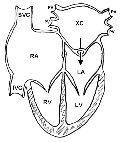
Synonyms: Subdivided left atrium, accessory atrium, heart with 3 atria, triatrial heart, cor triatriatum sinister, cor triatriatum sinistrum, cor triatriatum dexter, cor triatriatum dextrum.
Overview
Anatomy
Epidemiology and demographics
Natural history, Complications, and Prognosis
Causes
Differentiating Cor triatriatum from other Disorders
Diagnosis
Echocardiography is the primary method to diagnose a divided atrium. The transthoracic two dimensional echocardiography is usually aided by transesophageal echocardiogram for further evaluation. MRI provides a better spatial resolution and tissue contrast as compared to echocardiogram. MRI is said to have higher sensitivity than echocardiogram and angiography.[1]
History and Symptoms | Physical Examination | Laboratory Tests | Electrocardiogram | Chest X Ray | MRI | CT | Echocardiography | Other Imaging Findings | Other Diagnostic Studies
Treatment
Medical therapy is primarily symptomatic, definitive therapy being surgical.
Medical: Medical Therapy
Surgical: Surgery
Primary Prevention | Secondary Prevention | Cost-Effectiveness of Therapy | Future or Investigational Therapies
References
- ↑ [Masui et al. Abnormalities of the Pulmonary Veins: Evaluation with MR imaging and comparison with cardiac angiography and echocardiography. Radiology 1991; 181:645]
External Links
- Image at Yale University
- Diagram at University of Hawaii
- Description at University of Hawaii
- Echocardiogram at ecocardiografia.info
- Goldminer: Cor triatriatum
- Echocardiogram at ecocardiografia.info
- Cardiac MRI
- Cor triatriatum sinistrum: A pathomorphological image
- Angiography: Cor triatriatum dexter