Cardiac tamponade electrocardiogram: Difference between revisions
Jump to navigation
Jump to search
No edit summary |
No edit summary |
||
| Line 1: | Line 1: | ||
{{Cardiac tamponade}} | {{Cardiac tamponade}} | ||
{{CMG}}; '''Associate Editors-In-Chief:''' {{CZ}}; [[Varun Kumar]], M.B.B.S. | {{CMG}}; '''Associate Editors-In-Chief:''' {{CZ}}; [[Varun Kumar]], M.B.B.S. | ||
==Overview== | |||
==Electrocardiogram== | ==Electrocardiogram== | ||
EKG findings of [[cardiac tamponade]] are: | EKG findings of [[cardiac tamponade]] are: | ||
* [[Sinus tachycardia]] | * [[Sinus tachycardia]] | ||
* [[Electrical alternans]] (beat-to-beat alterations in the QRS complex due to swinging of heart in pericardial fluid) | * [[Electrical alternans]] (beat-to-beat alterations in the QRS complex due to the swinging of the heart in pericardial fluid) | ||
* Low voltage QRS complexes (Low QRS voltage is defined as maximum QRS amplitude in precordial lead < 1 mV and <0.5 mV in the limb leads due to insulating properties of fluid) <ref>Longmore, M., Wilkinson, I.B., Rajagopalan, S. (2004) (6th Ed.). Oxford Handbook of Clinical Medicine. Oxford: Oxford University Press ISBN 9780198568377 </ref>. | * Low voltage QRS complexes (Low QRS voltage is defined as maximum QRS amplitude in precordial lead < 1 mV and < 0.5 mV in the limb leads due to insulating properties of fluid) <ref>Longmore, M., Wilkinson, I.B., Rajagopalan, S. (2004) (6th Ed.). Oxford Handbook of Clinical Medicine. Oxford: Oxford University Press ISBN 9780198568377 </ref>. | ||
* [[ST segment]] <ref>Dolan, B., Holt, L. (2000). Accident & Emergency: Theory into practice. London: Bailliere Tindall ISBN 978-0702022395 </ref>. | * [[ST segment]] <ref>Dolan, B., Holt, L. (2000). Accident & Emergency: Theory into practice. London: Bailliere Tindall ISBN 978-0702022395 </ref>. | ||
* EKG findings of [[pericarditis]] | * EKG findings of [[pericarditis]] and [[pericardial effusion]] may be seen if these conditions are accompanying tamponade. | ||
<div align="left"> | <div align="left"> | ||
<gallery heights="250" widths="500"> | <gallery heights="250" widths="500"> | ||
Revision as of 18:35, 19 September 2012
|
Cardiac tamponade Microchapters |
|
Diagnosis |
|---|
|
Treatment |
|
Case Studies |
|
Cardiac tamponade electrocardiogram On the Web |
|
American Roentgen Ray Society Images of Cardiac tamponade electrocardiogram |
|
Risk calculators and risk factors for Cardiac tamponade electrocardiogram |
Editor-In-Chief: C. Michael Gibson, M.S., M.D. [1]; Associate Editors-In-Chief: Cafer Zorkun, M.D., Ph.D. [2]; Varun Kumar, M.B.B.S.
Overview
Electrocardiogram
EKG findings of cardiac tamponade are:
- Sinus tachycardia
- Electrical alternans (beat-to-beat alterations in the QRS complex due to the swinging of the heart in pericardial fluid)
- Low voltage QRS complexes (Low QRS voltage is defined as maximum QRS amplitude in precordial lead < 1 mV and < 0.5 mV in the limb leads due to insulating properties of fluid) [1].
- ST segment [2].
- EKG findings of pericarditis and pericardial effusion may be seen if these conditions are accompanying tamponade.
-
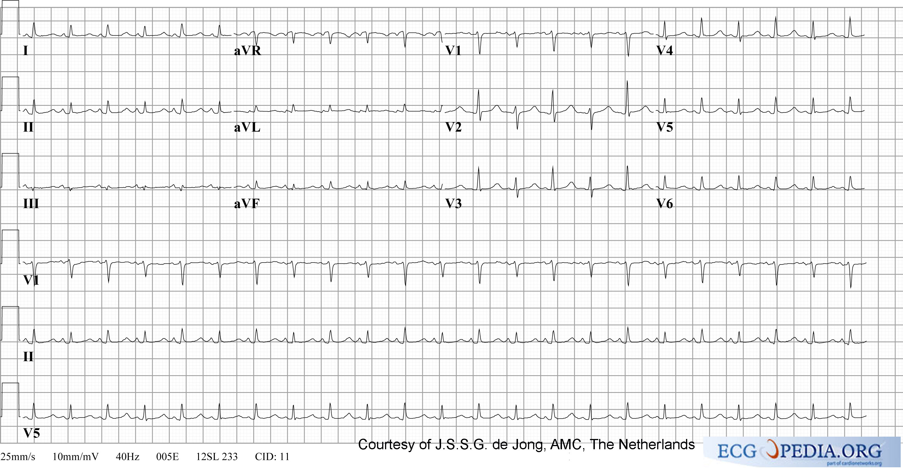
Cardiac Tamponade with low voltage QRS complex and Electrical Alternans
-
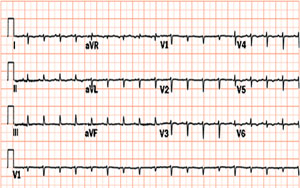
12 lead EKG shows Cardiac Tamponade with Electrical Alternans

