Atrial septal defect echocardiography sinus venosus

Revision as of 17:32, 21 August 2011 by Priyamvada Singh (talk | contribs)
Jump to navigation Jump to search

Atrial Septal Defect Microchapters

Home

Patient Information

Overview

Anatomy

Classification

Ostium Secundum Atrial Septal Defect
Ostium Primum Atrial Septal Defect
Sinus Venosus Atrial Septal Defect
Coronary Sinus
Patent Foramen Ovale
Common or Single Atrium

Pathophysiology

Epidemiology and Demographics

Risk Factors

Natural History and Prognosis

Complications

Diagnosis

History and Symptoms

Physical Examination

Electrocardiogram

Chest X Ray

CT

MRI

Echocardiography

Transesophageal Echocardiography
Transthoracic Echocardiography
Contrast Echocardiography
M-Mode
Doppler

Transcranial Doppler Ultrasound

Cardiac Catheterization

Exercise Testing

ACC/AHA Guidelines for Evaluation of Unoperated Patients

Treatment

Medical Therapy

Surgery

Indications for Surgical Repair
Surgical Closure
Minimally Invasive Repair


Robotic ASD Repair
Percutaneous Closure
Post-Surgical Follow Up

Special Scenarios

Pregnancy
Diving and Decompression Sickness
Paradoxical Emboli
Pulmonary Hypertension
Eisenmenger's Syndrome
Atmospheric Pressure

Case Studies

Case #1

Atrial septal defect echocardiography sinus venosus On the Web

Most recent articles

Most cited articles

Review articles

CME Programs

Powerpoint slides

Images

American Roentgen Ray Society Images of Atrial septal defect echocardiography sinus venosus

All Images
X-rays
Echo & Ultrasound
CT Images
MRI

Ongoing Trials at Clinical Trials.gov

US National Guidelines Clearinghouse

NICE Guidance

FDA on Atrial septal defect echocardiography sinus venosus

CDC on Atrial septal defect echocardiography sinus venosus

Atrial septal defect echocardiography sinus venosus in the news

Blogs on Atrial septal defect echocardiography sinus venosus

Directions to Hospitals Treating Type page name here

Risk calculators and risk factors for Atrial septal defect echocardiography sinus venosus

Editor-In-Chief: C. Michael Gibson, M.S., M.D. [1]; Eli V. Gelfand, MD; Keri Shafer, M.D. [2];

Associate Editors-In-Chief: Cafer Zorkun, M.D., Ph.D. [3]; Priyamvada Singh, MBBS [[4]]

Assistant Editor-In-Chief: Kristin Feeney, B.S. [[5]]

Overview

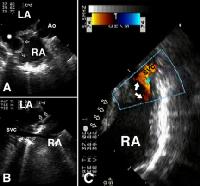
Echocardiography may be used as a diagnostic tool in the evaluation of an atrial septal defect. Common malformations of the septal wall include: ostium primum, ostium secundum, sinus venosus, and patent foramen ovale. Uncommonly, a defect may occur in the coronary sinus. Specific characteristics exist in echocardiography to identify these classifications of atrial septal defects.

Echocardiography and Sinus Venosus Defects

Characteristics of sinus venosus defects include:

  • Seen best in the subcostal four-chamber view by paying special attention to the superior and posterior portions of the atria.
  • Occurs at the top of the septum near the insertion of the superior vena cava.

<youtube v=dA2Zjq4Cx20/>

References

Useful Links

Yale Congenital Heart Disease- ASD [6]

Template:WH Template:WS