Aortic arch anomalies other imaging findings
|
Aortic arch anomalies Microchapters |
|
Diagnosis |
|---|
|
Treatment |
|
Case Studies |
|
Aortic arch anomalies other imaging findings On the Web |
|
American Roentgen Ray Society Images of Aortic arch anomalies other imaging findings |
|
Risk calculators and risk factors for Aortic arch anomalies other imaging findings |
Editor-In-Chief: C. Michael Gibson, M.S., M.D. [1]
Associate Editor-In-Chief: Cafer Zorkun, M.D., Ph.D. [2] Keri Shafer, M.D. [3] Priyamvada Singh, MBBS [[4]]
Assistant Editor-In-Chief: Kristin Feeney, B.S. [[5]]
Other Imaging Findings
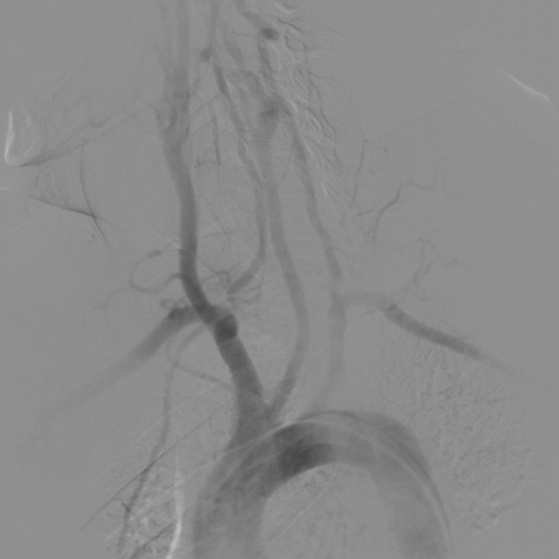
Aortography
Still aortographic images:
-
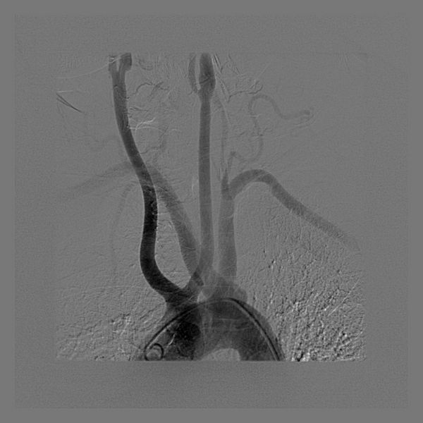
Subclavian Bovine Carotid
-
Bovine Aortic Arch


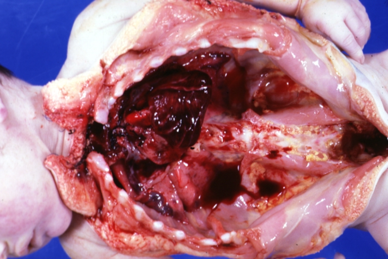
Anatomical Dissection

References
Additional Reading
- Moss and Adams' Heart Disease in Infants, Children, and Adolescents Hugh D. Allen, Arthur J. Moss, David J. Driscoll, Forrest H. Adams, Timothy F. Feltes, Robert E. Shaddy, 2007 ISBN 0781786843
- Hurst's the Heart, Fuster V, 12th ed. 2008, ISBN 978-0-07-149928-6
- Willerson JT, Cardiovascular Medicine, 3rd ed., 2007, ISBN 978-1-84628-188-4
External Links
Acknowledgements
The content on this page was first contributed by: C. Michael Gibson, M.S., M.D.