Adenoma
For patient information click here Template:DiseaseDisorder infobox
|
WikiDoc Resources for Adenoma |
|
Articles |
|---|
|
Most recent articles on Adenoma |
|
Media |
|
Evidence Based Medicine |
|
Clinical Trials |
|
Ongoing Trials on Adenoma at Clinical Trials.gov Clinical Trials on Adenoma at Google
|
|
Guidelines / Policies / Govt |
|
US National Guidelines Clearinghouse on Adenoma
|
|
Books |
|
News |
|
Commentary |
|
Definitions |
|
Patient Resources / Community |
|
Directions to Hospitals Treating Adenoma Risk calculators and risk factors for Adenoma
|
|
Healthcare Provider Resources |
|
Causes & Risk Factors for Adenoma |
|
Continuing Medical Education (CME) |
|
International |
|
|
|
Business |
|
Experimental / Informatics |
Editor-In-Chief: C. Michael Gibson, M.S., M.D. [1]
|
Adenoma Microchapters |
|
Diagnosis |
|---|
|
Treatment |
|
Case Studies |
|
Adenoma On the Web |
|
American Roentgen Ray Society Images of Adenoma |
Overview
An adenoma is a collection of growths (-oma) of glandular origin. Adenomas can grow from many organs including the colon, adrenal, pituitary, thyroid, etc. These growths are benign, although over time they may progress to become malignant (at which point they are called adenocarcinomas). Though adenomas are benign, they may unfortunately cause serious health complications by compressing other structures (mass effect) and by producing large amounts of hormones in an unregulated, non-feedback dependent manner (paraneoplastic syndrome).
Histopathology
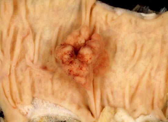
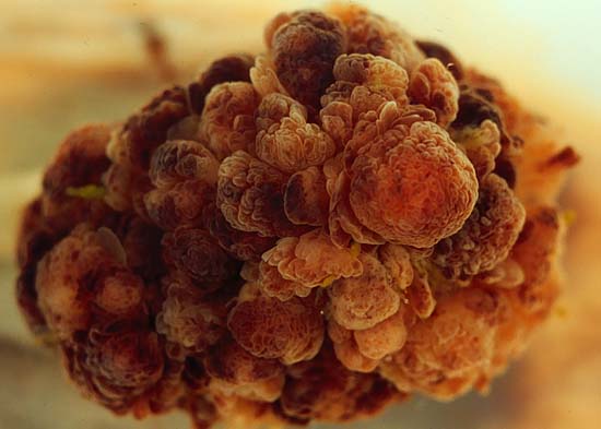
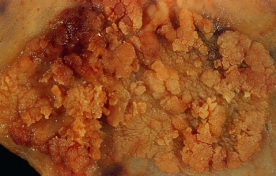
Adenoma is a benign epithelial tumor arising in epithelium of mucosa (stomach, small intestine and bowel), glands (endocrine and exocrine) and ducts. In hollow organs (digestive tract) the adenoma grows upwards into the lumen - adenomatous Polyp (or polypoid adenoma.
Depending on the type of the insertion base, adenoma may be pedunculated lobular head with a long slender stalk, covered by normal mucosa or sessile (broad base).
The adenomatous proliferation is characterized by different degrees of cell dysplasia (atypia or loss of normal differentiation of epithelium) irregular cells with hyperchromatic nuclei, (pseudo)stratified nuclei, nucleolus, decreased mucosecretion and mitosis. The architecture may be tubular, villous or tubulo-villous. Basement membrane and muscularis mucosae are intact.
-
Adenomatous polyp of the colon. Courtesy of Ed Uthman, MD
-
Tubulovillous polyp of the colon. Courtesy of Ed Uthman, MD
-
Villous adenoma of the colon. Courtesy of Ed Uthman, MD


Locations
Colon
Adenomas of the colon are quite prevalent. They are found commonly at colonoscopy. They are removed because of their tendency to become malignant and lead to colon cancer.
Renal
This is a tumor which is most often small and asymptomatic and its derived from renal tubules. It may be a precursor lesion to renal carcinoma.
Adrenal
Adrenal adenomas are common, 1 in 10 people have them malignant and asymptomatic. They are often found on of the abdomen, usually not as the focus of investigation; they are usually incidental findings. About one in 10,000 is malignant. Thus, a biopsy is rarely called for, especially if the lesion is homogeneous and smaller than 3 centimeters. Follow-up images in three to six months can confirm the stability of the growth.
While some adrenal adenomas do not secrete hormones at all , often some secrete cortisol causing Cushing's syndrome, aldosterone causing Conn's syndrome or androgens causing hyperandrogenism.
MRI
-
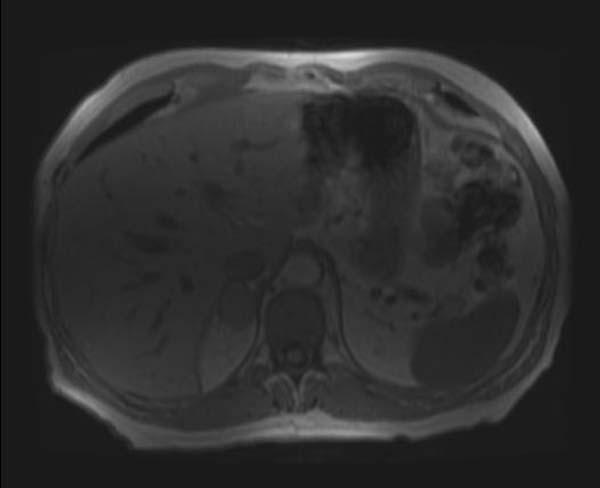
Adrenal adenoma
-
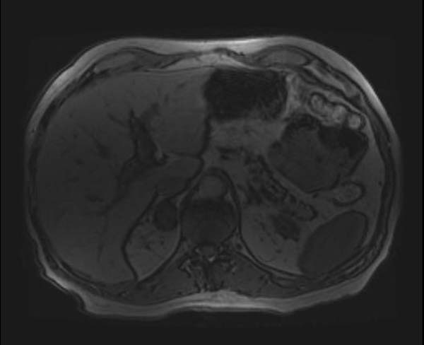
Adrenal adenoma
-
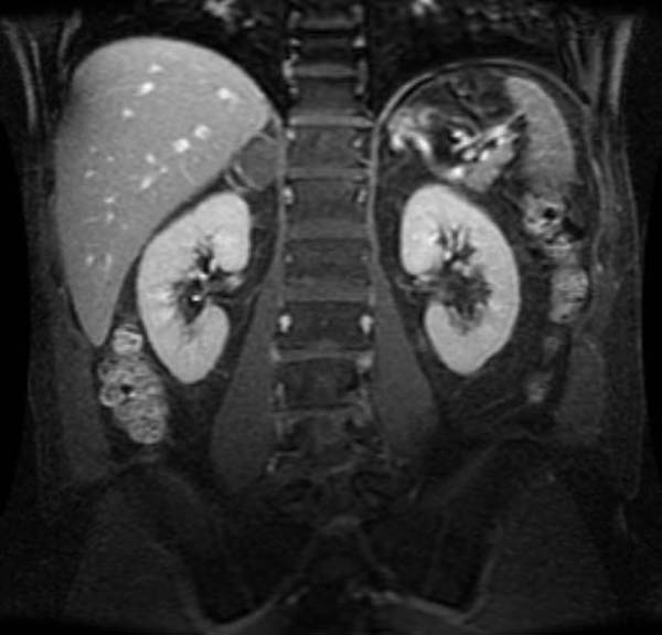
Adrenal adenoma


Thyroid
About one in 10 people are found to have solitary thyroid nodules. Investigation is required because a small percentage of these are malignant. Biopsy usually confirms the growth to be an adenoma, but sometimes, excision at surgery is required, especially when the cells found at biopsy are of the follicular type.
Pituitary
Pituitary adenomas are commonly seen in 10% of the neurological patients. A lot of them remain undiagnosed. Treatment is usually surgical, to which patients generally respond well. The most common subtype, prolactinoma, is seen more often in women, and is frequently diagnosed during pregnancy as the hormone progesterone increases its growth. Medical therapy bromocriptine generally suppresses prolactinomas; progesterone antagonist therapy has not proven to be successful.
Liver
Hepatocellular adenoma, Hepatic adenomas are a rare benign tumour of the liver, which may present with hepatomegaly or other symptoms.
Breast
Breast adenomas are called fibroadenomas. They are often very small and difficult to detect. Often there are no symptoms. Treatments can include a needle biopsy, and/or removal.
Appendix
Adenomas can also appear in the appendix. The condition is extremely rare and most physicians will never encounter an actual case, but they do happen. The most common version is called cystadenoma. They are usually discovered in the course of examination of the tissue following an appendectomy. If the appendix has ruptured and a tumor is present this presents challenges, especially if malignant cells have formed and thus spread to the abdomen. In 1995 Former Vice President Dan Quayle was found to have cystadenoma in the appendix.
Diagnosis
Computerized Tomography
Unenchanced CT
- Adrenal adenomas appear as small (<3 cm), well-defined homogeneous masses that are typically hypoattenuating relative to the liver.
- At an attenuation value of less than 0 HU at unenhanced CT, the diagnosis of an adenoma can be made with 100% confidence; however, this threshold has only 47% sensitivity.
- At cutoff of 18 HU, a diagnosis of adenoma was made with 100% specificity and 85% sensitivity, compared to the specificity:sensitivity ratio of 68%:100% with a more conservative cutoff of 10 HU.
- A rational approach advocated by some authorities is to choose the CT number threshold on the basis of the patient’s risk for metastatic disease. For example, a threshold of 10 HU could be applied to older patients or to those with known primary malignancies. A threshold of 18 HU could be applied to younger patients without underlying cancer.
Enhanced CT
- Lipid-poor adenomas are more difficult to diagnose because the CT numbers increase and approach those of soft tissue.
- Contrast-enhanced imaging with 10-minute-delayed CT scans may be helpful in these cases.
- By using a threshold of 30 HU, the sensitivity and specificity for delayed contrast-enhanced CT in the characterization of benign disease are 80% and 100%, respectively.
- A relative percentage washout of more than 50% in the delayed study represents a sensitivity and specificity of 98% and 100%, respectively, for the detection of adenoma.
Magnetic Resonance Imaging
- Chemical shift MR imaging can be used for further characterization when CT results are indeterminate.
- Because of the high sensitivity of chemical shift MR imaging to minute amounts of intravoxel fat, MR imaging demonstrates signal intensity loss on opposed-phase images in the majority of adenomas, with a sensitivity of 89% for lesions with an attenuation of 10–30 HU and 100% for lesions with an attenuation of 10–20 HU with a maintained specificity of 100%.
-

CT
-
T1 in phase
-
T1 out of phase
-

Template:Epithelial neoplasms
Template:SIB
da:Adenom de:Adenom gl:Adenoma it:Adenoma ms:Adenoma nl:Adenoom no:Adenom fi:Adenooma sv:Adenom uk:Аденома ur:ورم غدہ