Cryptogenic organizing pneumonia pathophysiology: Difference between revisions
Sergekorjian (talk | contribs) No edit summary |
|||
| (35 intermediate revisions by 3 users not shown) | |||
| Line 1: | Line 1: | ||
__NOTOC__ | __NOTOC__ | ||
{{Cryptogenic organizing pneumonia}} | {{Cryptogenic organizing pneumonia}} | ||
{{CMG}} {{AE}} {{MKK}} {{SSK}} | |||
==Overview== | ==Overview== | ||
[[Cryptogenic organizing pneumonia]] is an idiopathic diffuse [[interstitial lung disease]] that affects the distal [[bronchioles]], respiratory [[bronchioles]], [[alveolar ducts]], and [[alveolar]] walls. The injury occurs within the [[alveolar]] wall. There is proliferation of [[granulation tissue]] which involves [[alveolar ducts]] and [[alveoli]]. There are 4 phases lead to the formation of mature fibrotic bud. | |||
==Pathophysiology== | ==Pathophysiology== | ||
Cryptogenic organizing pneumonia | |||
===Pathogenesis=== | |||
[[Cryptogenic organizing pneumonia]] is caused by excessive proliferation of [[granulation tissue]] which involves [[alveolar ducts]] and [[alveoli]]. [[Granulation tissue]] may extend from one [[alveolus]] to the adjacent one leading to the formation of mature fibrotic bud which gives characteristic "butterfly" pattern. | |||
Various phases involved in the pathogenesis of [[cryptogenic organizing pneumonia]] are:<ref name="pmid11790668">{{cite journal |vauthors= |title=American Thoracic Society/European Respiratory Society International Multidisciplinary Consensus Classification of the Idiopathic Interstitial Pneumonias. This joint statement of the American Thoracic Society (ATS), and the European Respiratory Society (ERS) was adopted by the ATS board of directors, June 2001 and by the ERS Executive Committee, June 2001 |journal=Am. J. Respir. Crit. Care Med. |volume=165 |issue=2 |pages=277–304 |date=January 2002 |pmid=11790668 |doi=10.1164/ajrccm.165.2.ats01 |url=}}</ref> | |||
1) '''Injury phase''' - The early phase of [[cryptogenic organizing pneumonia]]. | |||
*It is characterized by the deposition of [[plasma proteins]] in the [[alveolar]] lumen. | |||
*Mechanism of early phase is an imbalance between [[coagulation]] and [[fibrinolytic]] cascade and activation of [[coagulation]] process which leads to [[fibrin]] deposition.<ref name="pmid16880372">{{cite journal |vauthors=Cordier JF |title=Cryptogenic organising pneumonia |journal=Eur. Respir. J. |volume=28 |issue=2 |pages=422–46 |date=August 2006 |pmid=16880372 |doi=10.1183/09031936.06.00013505 |url=}}</ref> | |||
2) '''Proliferating phase''' - The second stage of the [[cryptogenic organizing pneumonia]] in which there is a formation of fibro-[[inflammatory]] buds. | |||
* [[Macrophages]] and [[inflammatory cells]] help in fragmentation of [[fibrin]]. | |||
* Activated [[fibroblasts]] differentiate into [[myofibroblasts]] which are migrating through gaps of the [[basal lamina]]. | |||
* [[Inflammatory cells]] and [[fibrin]] are progressively replaced by aggregated [[fibroblasts]]/[[myofibroblasts]] intermixed with a [[Loose connective tissue|loose connective]] matrix tissue rich in [[collagen]] (especially collagen I), [[fibronectin]], [[procollagen]] type III and [[proteoglycans]]. | |||
* [[Alveolar]] [[epithelial cells]] proliferate, restoring the continuity of the [[Alveolar-capillary barrier|alveolar-capillary]] membrane and the integrity of the [[alveolar]] unit. | |||
3) '''Mature phase''' - The third stage is characterized by the formation of mature fibrotic buds which gives characteristic "butterfly" pattern. | |||
*In [[alveolar]] buds, there are [[myofibroblasts]], organized in [[concentric]] rings alternating with layers of [[collagen]] bundles. | |||
4)'''Resolution phase''' - The fourth stage, this stage usually resolves if there is the preservation of [[alveolar]] basal laminae. | |||
==Associated Conditions== | |||
[[Cryptogenic organizing pneumonia]] is associated with the following conditions:<ref name="urlOrganising pneumonia | Thorax">{{cite web |url=http://thorax.bmj.com/content/55/4/318#ref-3 |title=Organising pneumonia | Thorax |format= |work= |accessdate=}}</ref><ref name="pmid7890282">{{cite journal |vauthors=Kwon KY, Myers JL, Swensen SJ, Colby TV |title=Middle lobe syndrome: a clinicopathological study of 21 patients |journal=Hum. Pathol. |volume=26 |issue=3 |pages=302–7 |date=March 1995 |pmid=7890282 |doi= |url=}}</ref> | |||
*Infectious [[pneumonia]] | |||
*[[Lung abscess]] | |||
*[[Empyema]] | |||
*[[Lung cancer]] | |||
*Chronic [[pulmonary fibrosis]] | |||
*[[Aspiration pneumonia]] | |||
*[[Acute respiratory distress syndrome|Adult respiratory distress syndrome]] | |||
*[[Pulmonary infarction]] | |||
*Middle lobe syndrome | |||
==Gross Pathology== | |||
On gross pathology of [[cryptogenic organizing pneumonia]], following features are seen: | |||
*There is the firm area with preservation of [[lung]] pattern which extends to thickened [[Pleurae|pleura]].<ref name="urlOrganising pneumonia | Thorax">{{cite web |url=http://thorax.bmj.com/content/55/4/318 |title=Organising pneumonia | Thorax |format= |work= |accessdate=}}</ref> | |||
==Microscopic Pathology== | |||
On microscopic histopathological analysis:<ref name="urlCryptogenic organising pneumonia | Radiology Reference Article | Radiopaedia.org">{{cite web |url=https://radiopaedia.org/articles/cryptogenic-organising-pneumonia-1 |title=Cryptogenic organising pneumonia | Radiology Reference Article | Radiopaedia.org |format= |work= |accessdate=}}</ref><ref name="pmid29404167">{{cite journal |vauthors=Akyıl FT, Ağca M, Mısırlıoğlu A, Arsev AA, Akyıl M, Sevim T |title=Organizing Pneumonia as a Histopathological Term |journal=Turk Thorac J |volume=18 |issue=3 |pages=82–87 |date=July 2017 |pmid=29404167 |pmc=5783087 |doi=10.5152/TurkThoracJ.2017.16047 |url=}}</ref> | |||
*It is characterized by chronic mild interstitial [[inflammation]] without [[fibrosis]]. | |||
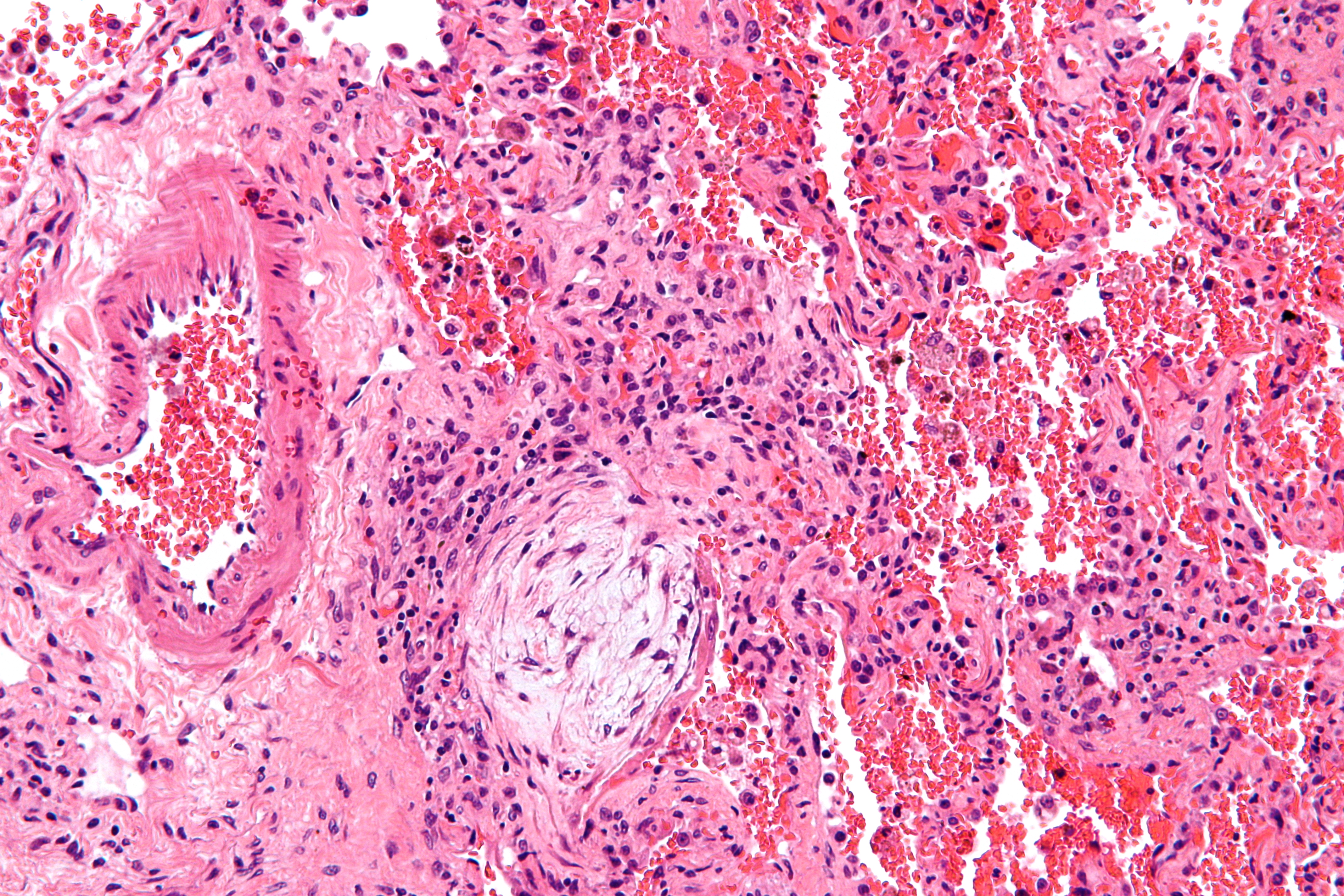
*There is the formation of buds of [[granulation tissue]] which is made of [[fibrous tissue]] (Masson bodies), [[mononuclear cells]] and foamy [[macrophages]], in the distal airspaces which cause [[secondary]] bronchiolar [[occlusion]] due to the presence of the [[inflammatory process]]. | |||
[[File:Masson body - high mag.jpg|200px|thumb|centre| source:By Nephron<ref>https://commons.wikimedia.org/w/index.php?curid=17325428</ref>]] | |||
==References== | ==References== | ||
{{Reflist|2}} | {{Reflist|2}} | ||
[[Category:Pulmonology]] | [[Category:Pulmonology]] | ||
{{WH}} | {{WH}} | ||
{{WS}} | {{WS}} | ||
Latest revision as of 14:16, 28 March 2018
|
Cryptogenic Organizing Pneumonia Microchapters |
|
Differentiating Cryptogenic organizing pneumonia from other Diseases |
|---|
|
Diagnosis |
|
Treatment |
|
Case Studies |
|
Cryptogenic organizing pneumonia pathophysiology On the Web |
|
American Roentgen Ray Society Images of Cryptogenic organizing pneumonia pathophysiology |
|
Cryptogenic organizing pneumonia pathophysiology in the news |
|
Directions to Hospitals Treating Cryptogenic organizing pneumonitis |
|
Risk calculators and risk factors for Cryptogenic organizing pneumonia pathophysiology |
Editor-In-Chief: C. Michael Gibson, M.S., M.D. [1] Associate Editor(s)-in-Chief: Manpreet Kaur, MD [2] Serge Korjian M.D.
Overview
Cryptogenic organizing pneumonia is an idiopathic diffuse interstitial lung disease that affects the distal bronchioles, respiratory bronchioles, alveolar ducts, and alveolar walls. The injury occurs within the alveolar wall. There is proliferation of granulation tissue which involves alveolar ducts and alveoli. There are 4 phases lead to the formation of mature fibrotic bud.
Pathophysiology
Pathogenesis
Cryptogenic organizing pneumonia is caused by excessive proliferation of granulation tissue which involves alveolar ducts and alveoli. Granulation tissue may extend from one alveolus to the adjacent one leading to the formation of mature fibrotic bud which gives characteristic "butterfly" pattern.
Various phases involved in the pathogenesis of cryptogenic organizing pneumonia are:[1]
1) Injury phase - The early phase of cryptogenic organizing pneumonia.
- It is characterized by the deposition of plasma proteins in the alveolar lumen.
- Mechanism of early phase is an imbalance between coagulation and fibrinolytic cascade and activation of coagulation process which leads to fibrin deposition.[2]
2) Proliferating phase - The second stage of the cryptogenic organizing pneumonia in which there is a formation of fibro-inflammatory buds.
- Macrophages and inflammatory cells help in fragmentation of fibrin.
- Activated fibroblasts differentiate into myofibroblasts which are migrating through gaps of the basal lamina.
- Inflammatory cells and fibrin are progressively replaced by aggregated fibroblasts/myofibroblasts intermixed with a loose connective matrix tissue rich in collagen (especially collagen I), fibronectin, procollagen type III and proteoglycans.
- Alveolar epithelial cells proliferate, restoring the continuity of the alveolar-capillary membrane and the integrity of the alveolar unit.
3) Mature phase - The third stage is characterized by the formation of mature fibrotic buds which gives characteristic "butterfly" pattern.
- In alveolar buds, there are myofibroblasts, organized in concentric rings alternating with layers of collagen bundles.
4)Resolution phase - The fourth stage, this stage usually resolves if there is the preservation of alveolar basal laminae.
Associated Conditions
Cryptogenic organizing pneumonia is associated with the following conditions:[3][4]
- Infectious pneumonia
- Lung abscess
- Empyema
- Lung cancer
- Chronic pulmonary fibrosis
- Aspiration pneumonia
- Adult respiratory distress syndrome
- Pulmonary infarction
- Middle lobe syndrome
Gross Pathology
On gross pathology of cryptogenic organizing pneumonia, following features are seen:
Microscopic Pathology
On microscopic histopathological analysis:[5][6]
- It is characterized by chronic mild interstitial inflammation without fibrosis.
- There is the formation of buds of granulation tissue which is made of fibrous tissue (Masson bodies), mononuclear cells and foamy macrophages, in the distal airspaces which cause secondary bronchiolar occlusion due to the presence of the inflammatory process.

References
- ↑ "American Thoracic Society/European Respiratory Society International Multidisciplinary Consensus Classification of the Idiopathic Interstitial Pneumonias. This joint statement of the American Thoracic Society (ATS), and the European Respiratory Society (ERS) was adopted by the ATS board of directors, June 2001 and by the ERS Executive Committee, June 2001". Am. J. Respir. Crit. Care Med. 165 (2): 277–304. January 2002. doi:10.1164/ajrccm.165.2.ats01. PMID 11790668.
- ↑ Cordier JF (August 2006). "Cryptogenic organising pneumonia". Eur. Respir. J. 28 (2): 422–46. doi:10.1183/09031936.06.00013505. PMID 16880372.
- ↑ 3.0 3.1 "Organising pneumonia | Thorax".
- ↑ Kwon KY, Myers JL, Swensen SJ, Colby TV (March 1995). "Middle lobe syndrome: a clinicopathological study of 21 patients". Hum. Pathol. 26 (3): 302–7. PMID 7890282.
- ↑ "Cryptogenic organising pneumonia | Radiology Reference Article | Radiopaedia.org".
- ↑ Akyıl FT, Ağca M, Mısırlıoğlu A, Arsev AA, Akyıl M, Sevim T (July 2017). "Organizing Pneumonia as a Histopathological Term". Turk Thorac J. 18 (3): 82–87. doi:10.5152/TurkThoracJ.2017.16047. PMC 5783087. PMID 29404167.
- ↑ https://commons.wikimedia.org/w/index.php?curid=17325428