Thyroid cartilage
Template:Infobox Anatomy Editor-In-Chief: C. Michael Gibson, M.S., M.D. [1]
The thyroid cartilage is the largest of the nine cartilages that make up the laryngeal skeleton, the cartilage structure in and around the trachea that contains the larynx.
Structure
It is composed of two plate-like laminae that come together on the anterior side of the cartilage to form a peak, called the laryngeal prominence. This prominence is often referred to as the "Adam's apple". The laryngeal prominence is obvious in both sexes, but it tends to be somewhat more robust in the adult male.
The lip of the thyroid cartilage just superior to the laryngeal prominence is called the thyroid notch or superior thyroid notch.
Layers and articulations
The two laminae that make up the main lateral, surfaces of the thyroid cartilage extend obliquely to cover either side of the trachea.
The posterior edge of each lamina articulates with the cricoid cartilage inferiorly at a joint called the cricothyroid joint.
Movement of the cartilage at this joint produces a change in tension at the vocal folds, which in turn produces variation in voice.
The entire superior edge of the thyroid cartilage is attached to the hyoid bone by the hyothyroid membrane.
Function
The thyroid cartilage forms the bulk of the anterior wall of the larynx, and serves to protect the vocal folds ("vocal cords") which are located directly behind it.
It also serves as an attachment for several laryngeal muscles.
Additional images
-
 Larynx
 -
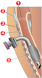 Tracheotomy neck profile
 -
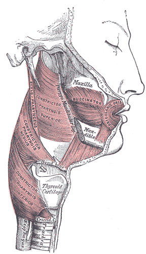 Muscles of the pharynx and cheek.
 -
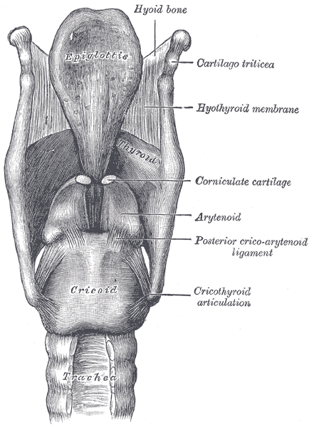
 The cartilages of the larynx. Posterior view.
 -
 Ligaments of the larynx. Posterior view.
 -
 Sagittal section of the larynx and upper part of the trachea.
 -
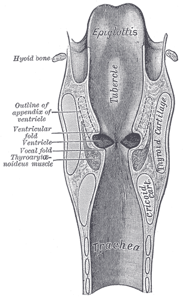 Coronal section of larynx and upper part of trachea.
 -
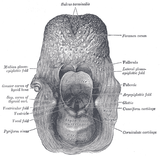 The entrance to the larynx, viewed from behind.
 -
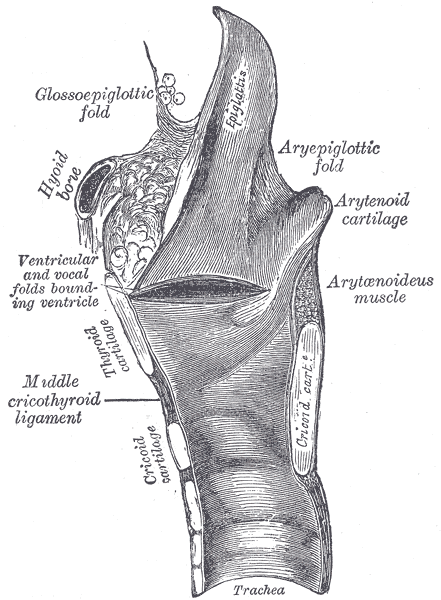
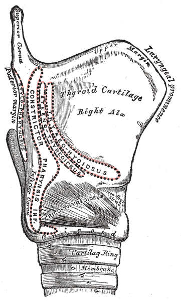 Side view of the larynx, showing muscular attachments.
 -
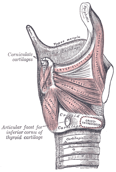 Muscles of larynx. Side view. Right lamina of thyroid cartilage removed.
 -
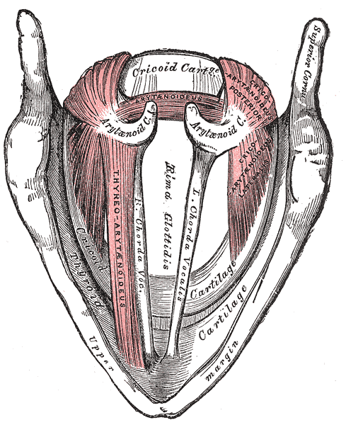 Muscles of the larynx, seen from above.
 -
 Front view of neck.
 -
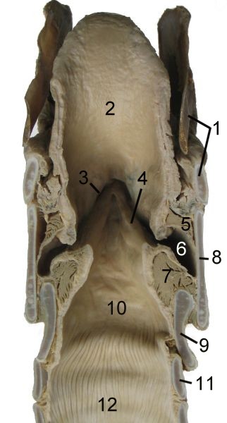 Cut through the larynx of a horse


