Superior ulnar collateral artery
Jump to navigation
Jump to search
Editor-In-Chief: C. Michael Gibson, M.S., M.D. [1]
The superior ulnar collateral artery (inferior profunda artery), of small size, arises from the brachial a little below the middle of the arm; it frequently springs from the upper part of the a. profunda brachii.
It pierces the medial intermuscular septum, and descends on the surface of the medial head of the Triceps brachii to the space between the medial epicondyle and olecranon, accompanied by the ulnar nerve, and ends under the Flexor carpi ulnaris by anastomosing with the posterior ulnar recurrent, and inferior ulnar collateral.
It sometimes sends a branch in front of the medial epicondyle, to anastomose with the anterior ulnar recurrent.
Additional images
-
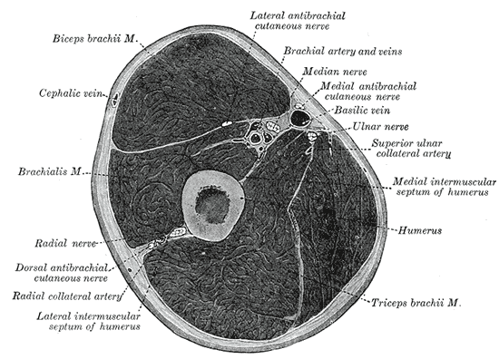 Cross-section through the middle of upper arm.
 -
 The brachial artery.

