Secondary bronchus
Jump to navigation
Jump to search
Editor-In-Chief: C. Michael Gibson, M.S., M.D. [1]
Overview
Secondary bronchi (also known as lobar bronchi) arise from the primary bronchi, with each one serving as the airway to a specific lobe of the lung.
Structure
They have relatively large lumens that are lined by respiratory epithelium. There is a smooth muscle layer below the epithelium arranged as two ribbons of muscle that spiral in opposite directions. This smooth muscle layer contains seromucous glands. Irregularly arranged plates of hyaline cartilage surround the smooth muscle. These plates give structural support to the bronchus and maintain the patency of the lumen.
Secondary bronchi of left lung
- superior lobe bronchus
- inferior lobe bronchus
Secondary bronchi of right lung
- superior lobe bronchus (or eparterial bronchus)
- middle lobe bronchus
- inferior lobe bronchus
Additional images
-
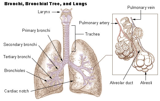 Bronchi, bronchial tree, and lungs
 -
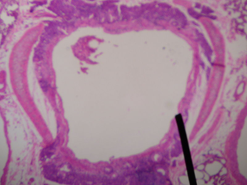 Cross sectional cut of a human secondary bronchus


References
- Gartner, Leslie P. and James L. Hiatt. Color Atlas of Histology, 3rd ed. (2000). ISBN 0-7817-3509-2
- Gartner, Leslie P. and James L. Hiatt. Color Textbook of Histology, 2nd ed. (2001). ISBN 0-7216-8806-3
External links
- Template:SUNYAnatomyFigs - "The divisions of the bronchus."
- Template:GPnotebook