Left lung
|
WikiDoc Resources for Left lung |
|
Articles |
|---|
|
Most recent articles on Left lung |
|
Media |
|
Evidence Based Medicine |
|
Clinical Trials |
|
Ongoing Trials on Left lung at Clinical Trials.gov Clinical Trials on Left lung at Google
|
|
Guidelines / Policies / Govt |
|
US National Guidelines Clearinghouse on Left lung
|
|
Books |
|
News |
|
Commentary |
|
Definitions |
|
Patient Resources / Community |
|
Patient resources on Left lung Discussion groups on Left lung Directions to Hospitals Treating Left lung Risk calculators and risk factors for Left lung
|
|
Healthcare Provider Resources |
|
Causes & Risk Factors for Left lung |
|
Continuing Medical Education (CME) |
|
International |
|
|
|
Business |
|
Experimental / Informatics |
Editor-In-Chief: C. Michael Gibson, M.S., M.D. [1]
Overview
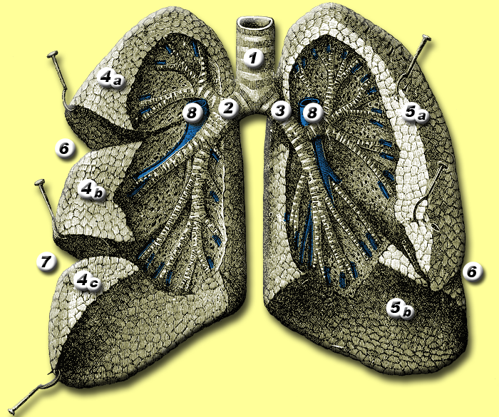
The Left lung is divided into two lobes, an upper and a lower, by the oblique fissure, which extends from the costal to the mediastinal surface of the lung both above and below the hilus.
As seen on the surface, this fissure begins on the mediastinal surface of the lung at the upper and posterior part of the hilus, and runs backward and upward to the posterior border, which it crosses at a point about 6 cm. below the apex.
It then extends downward and forward over the costal surface, and reaches the lower border a little behind its anterior extremity, and its further course can be followed upward and backward across the mediastinal surface as far as the lower part of the hilus.
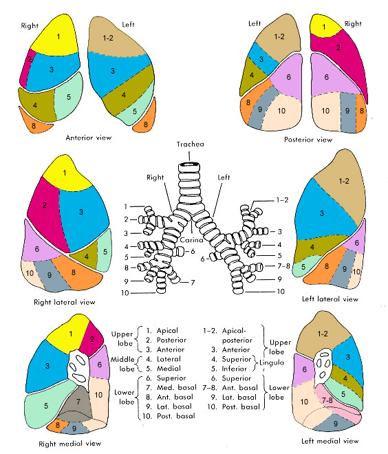
Lobes
The superior lobe lies above and in front of this fissure, and includes the apex, the anterior border, and a considerable part of the costal surface and the greater part of the mediastinal surface of the lung.
The inferior lobe, the larger of the two, is situated below and behind the fissure, and comprises almost the whole of the base, a large portion of the costal surface, and the greater part of the posterior border.
Impressions
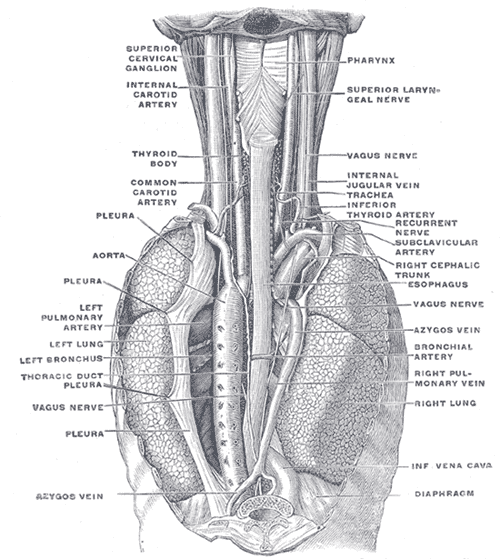
On the mediastinal surface, immediately above the hilus, is a well-marked curved furrow produced by the aortic arch, and running upward from this toward the apex is a groove accommodating the left subclavian artery; a slight impression in front of the latter and close to the margin of the lung lodges the left innominate vein.
Behind the hilus and pulmonary ligament is a vertical furrow produced by the descending aorta, and in front of this, near the base of the lung, the lower part of the esophagus causes a shallow impression.
Additional images
-
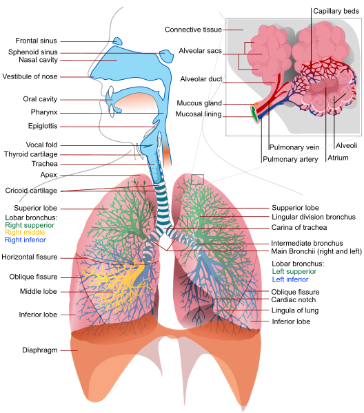
diagram of the respiratory system
-
Anatomy of lungs.
-

Front view of heart and lungs.
-

Transverse section of thorax, showing relations of pulmonary artery.
-
The position and relation of the esophagus in the cervical region and in the posterior mediastinum. Seen from behind.
-
The thymus of a full-time fetus, exposed in situ.


See also
External links
- Template:Chorus
- Template:SUNYAnatomyFigs - "Mediastinal surface of the left lung."
- Diagram and quiz at cancer.gov
- Slide at mgccc.cc.ms.us
- Template:RocheLexicon
- Template:GPnotebook - "lobes (left lung)"
- Diagram at md.chula.ac.th