Rectus femoris muscle
Overview
The Rectus femoris muscle is one of the four quadriceps muscles of the human body. (The others are the vastus medialis, the vastus intermedius (deep to the rectus femoris), and the vastus lateralis. All four combine to form the quadriceps tendon, which inserts into the patella and continues as the patellar ligament.)
The Rectus femoris is situated in the middle of the front of the thigh; it is fusiform in shape, and its superficial fibers are arranged in a bipenniform manner, the deep fibers running straight down to the deep aponeurosis.
Origin and insertion
It arises by two tendons: one, the anterior or straight, from the anterior inferior iliac spine; the other, the posterior or reflected, from a groove above the brim of the acetabulum.
The two unite at an acute angle, and spread into an aponeurosis which is prolonged downward on the anterior surface of the muscle, and from this the muscular fibers arise.
The muscle ends in a broad and thick aponeurosis which occupies the lower two-thirds of its posterior surface, and, gradually becoming narrowed into a flattened tendon, is inserted into the base of the patella.
Functions
- Hip Flexion
- Knee Extension
The rectus femoris is the only muscle in the quadriceps group that is involved in hip flexion, since it is the only one that originates in the pelvis and not the femur.
The rectus femoris is a weaker hip flexor when the knee is extended because it is already shortened and thus suffers from active insufficiency. In essence: the action of raising a straightened leg will recruit more iliacus, psoas major, tensor fasciae latae, and the remaining hip flexors than it will the rectus femoris.
Similarly, the rectus femoris is not dominant in knee extension when the hip is flexed since it is already shortened and thus suffers from active insufficiency. In essence: the action of extending a leg from a seated position is primarily driven by the vastus lateralis, vastus medialis, and vastus intermedius, and less by the rectus femoris.
The rectus femoris is considered a direct antagonist to the hamstrings. The hamstrings oppkjuose the rectus femoris at the hip joint through extension and at the knee joint through flexion. Simultaneous contraction of the rectus femoris and hamstrings results in simultaneous hip and knee extension due to leverage (Lombard's Paradox.)
Additional images
-
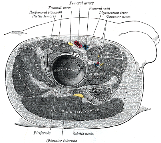 Structures surrounding right hip-joint.
 -
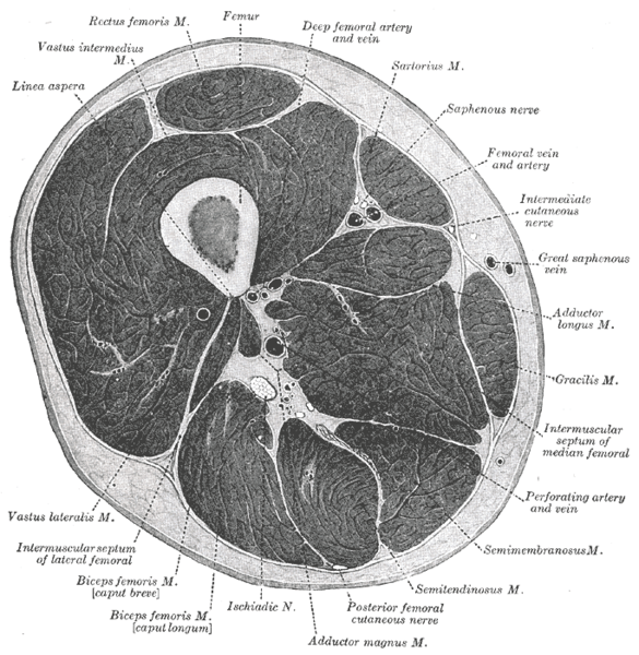 Cross-section through the middle of the thigh.
 -
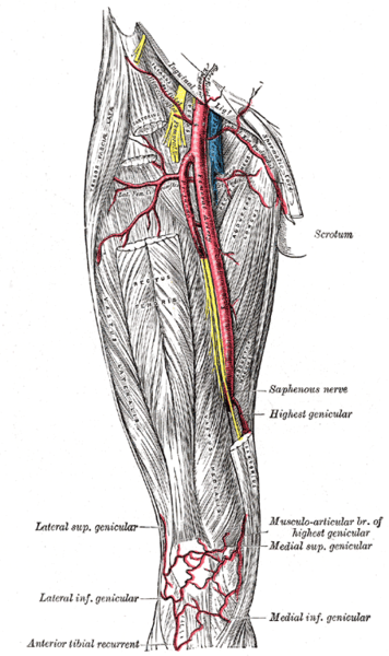 The femoral artery.
 -
 Nerves of the right lower extremity. Front view.


