Pericardial effusion pathophysiology
|
Pericardial effusion Microchapters |
|
Diagnosis |
|---|
|
Treatment |
|
Case Studies |
|
Pericardial effusion pathophysiology On the Web |
|
American Roentgen Ray Society Images of Pericardial effusion pathophysiology |
|
Risk calculators and risk factors for Pericardial effusion pathophysiology |
Editor-In-Chief: C. Michael Gibson, M.S., M.D. [1]; Associate Editor-In-Chief: Abdelrahman Ibrahim Abushouk, MD[2], Varun Kumar, M.B.B.S.
Overview
Pericardial effusion usually results from a disturbed equilibrium between the production and reabsorption of pericardial fluid. This can occur in infections and inflammations where there is increased production of pericardial fluid, increased microvascular pressure as in cardiac failure and renal failure cause, decreased plasma oncotic pressure as in cirrhosis and nephrotic syndrome, or in malignancy and hypothyroidism where there is inadequate drainage of the fluid.
Pathophysiology
Physiology
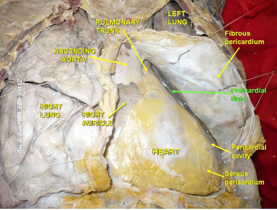
- Pericardium surrounds the heart and it consists of two layers, parietal and visceral layers [1].
- The space between the layers is known as the pericardial cavity.
- It usually contains small amount of fluid, approximately 15-50ml, which acts as a lubricating agent between the layers.
- This fluid enters the pericardial space from the capillaries into the visceral pericardium.
- This fluid is drained by lymphatics [2].
- When this fluid production-drainage mechanism is altered, excess fluid accumulates in the pericardial cavity and this is referred to as pericardial effusion.

Pathogenesis
Therefore, pericardial effusion occurs when there is:
- Increased capillary membrane permeability: Infection or inflammation may lead to exudative fluid or hemorrhagic effusion which have high protein levels. The pericardial effusion observed in the following conditions results from increased permeability of the capillary membrane [4].
- Viral/bacterial infections such as adenovirus infection and tuberculosis
- Autoimmune diseases such as sarcoidosis, SLE and rheumatoid arthritis
- Penetrating trauma which injure the blood vessels and cause hemorrhage into the pericardial space
- Malignancies such as pulmonary carcinoma may metastasize to the pericardium and can disrupt pericardial anatomy and vasculature
- Increased microvascular pressure: Hypervolemic states like cardiac failure and renal failure cause pericardial effusion due to increased microvascular pressure [5].
- Decreased plasma oncotic pressure: Pericardial effusion seen in cirrhosis and nephrotic syndrome is due to decreased plasma oncotic pressure secondary to hypoalbuminemia.
- Decreased drainage of pericardial fluid: Pericardial effusion may occur in malignancies and hypothyroidism due to decreased drainage of the pericardial fluid by the lymphatics [6][7].
Genetics
There are no known genetic causes of pericardial effusion.
Associated Conditions
Conditions associated with pericardial effusion include:
- Other pericardial diseases e.g. pericarditis.
- Parapneumonic effusion.
- Autoimmune disorders, such as rheumatoid arthritis or lupus.
- Malignancy
Gross Pathology
On gross pathology, enlarged cardiac cavity, compressed cardiac chambers (with large effusions), heart swinging within the effusion fluid are characteristic findings of pericardial effusion. Further, the color of the effusion fluid may give an insight into the possible effusion cause.
Microscopic Pathology
On microscopic histopathological analysis, there are no characteristic features of pericardial effusion. However, analysis of the pericardial fluid itself may give insights into the underlying cause e.g. numerous pus cells would indicate a pyogenic/inflammatory cause, lymphocytes indicate viral infection, and malignant cells indicate malignant seeding into the pericardium.
References
- ↑ Hoit BD (2017). "Anatomy and Physiology of the Pericardium". Cardiol Clin. 35 (4): 481–490. doi:10.1016/j.ccl.2017.07.002. PMID 29025540.
- ↑ Rodriguez ER, Tan CD (2017). "Structure and Anatomy of the Human Pericardium". Prog Cardiovasc Dis. 59 (4): 327–340. doi:10.1016/j.pcad.2016.12.010. PMID 28062264.
- ↑ https://commons.wikimedia.org/wiki/File:Slide14gggg.JPG/
- ↑ Vakamudi S, Ho N, Cremer PC (2017). "Pericardial Effusions: Causes, Diagnosis, and Management". Prog Cardiovasc Dis. 59 (4): 380–388. doi:10.1016/j.pcad.2016.12.009. PMID 28062268.
- ↑ Patel Y, Agarwal V, Argulian E (2018). "Relation of Blood Pressure to Severity of Pericardial Effusion". Am J Cardiol. 121 (11): 1409–1412. doi:10.1016/j.amjcard.2018.02.023. PMID 29580632.
- ↑ Scardulla F, Rinaudo A, Pasta S, Scardulla C (2015). "Mechanics of pericardial effusion: a simulation study". Proc Inst Mech Eng H. 229 (3): 205–14. doi:10.1177/0954411915574012. PMID 25833996.
- ↑ Refaat MM, Katz WE (2011). "Neoplastic pericardial effusion". Clin Cardiol. 34 (10): 593–8. doi:10.1002/clc.20936. PMC 6652358 Check
|pmc=value (help). PMID 21928406.