Parotid duct
Template:Infobox Anatomy Editor-In-Chief: C. Michael Gibson, M.S., M.D. [1]
Overview
The parotid duct is also known as koslo's duct. Saliva from the parotid gland passes through it to the mouth.
It pierces the buccal fat, buccopharyngeal fascia and buccinator muscle then opens into the vestibule of the mouth opposite the upper 2nd molar tooth. The buccinator acts as a valve which prevents inflation of the duct during blowing. Running along with the duct superiorly is the transverse facial artery and upper buccal nerve, inferiorly is the lower buccal nerve.
Pathology
Blockage, whether caused by salivary duct stones or external compression, may cause pain and swelling of the parotid gland (parotitis)
Eponym
It is named after Niels Stensen (1638-1686), a Danish anatomist credited with its discovery.
Additional images
-
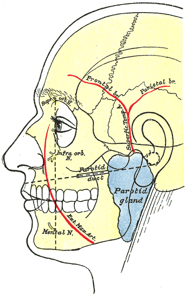 Outline of side of face, showing chief surface markings.
 -
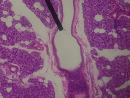 interlobular duct.
 -
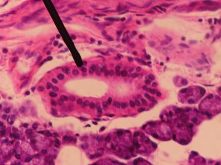 striated duct.


External links
- Diagram at MSU
- ent/178 at eMedicine - Parotid duct injuries
- Template:WhoNamedIt
Template:Digestive-stub Template:Head and neck general