Paragonimus infection pathophysiology
|
Paragonimus infection Microchapters |
|
Diagnosis |
|---|
|
Treatment |
|
Case Studies |
|
Paragonimus infection pathophysiology On the Web |
|
American Roentgen Ray Society Images of Paragonimus infection pathophysiology |
|
Risk calculators and risk factors for Paragonimus infection pathophysiology |
Editor-In-Chief: C. Michael Gibson, M.S., M.D. [1]
Pathophysiology

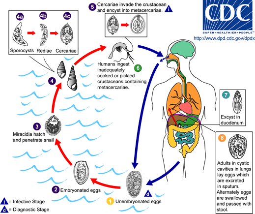
The eggs are excreted unembryonated in the sputum, or alternately they are swallowed and passed with stool.
In the external environment, the eggs become embryonated, and miracidia hatch and seek the first intermediate host, a snail, and penetrate its soft tissues.
Miracidia go through several developmental stages inside the snail: sporocysts, rediae, with the latter giving rise to many cercariae, which emerge from the snail.
The cercariae invade the second intermediate host, a crustacean such as a crab or crayfish, where they encyst and become metacercariae. This is the infective stage for the mammalian host.
Human infection with P. westermani occurs by eating inadequately cooked or pickled crab or crayfish that harbor metacercariae of the parasite. The metacercariae excyst in the duodenum, penetrate through the intestinal wall into the peritoneal cavity, then through the abdominal wall and diaphragm into the lungs, where they become encapsulated and develop into adults (7.5 to 12 mm by 4 to 6 mm).
The worms can also reach other organs and tissues, such as the brain and striated muscles, respectively. However, when this takes place completion of the life cycles is not achieved, because the eggs laid cannot exit these sites. Time from infection to oviposition is 65 to 90 days.
Infections may persist for 20 years in humans. Animals such as pigs, dogs, and a variety of feline species can also harbor P. westermani.