Meckel's cartilage
Template:Infobox Embryology Editor-In-Chief: C. Michael Gibson, M.S., M.D. [1]
The cartilaginous bar of the mandibular arch is formed by what are known as Meckel’s cartilages (right and left) ; above this the incus is developed.
The dorsal end of each cartilage is connected with the ear-capsule and is ossified to form the malleus; the ventral ends meet each other in the region of the symphysis menti, and are usually regarded as undergoing ossification to form that portion of the mandible which contains the incisor teeth.
The intervening part of the cartilage disappears; the portion immediately adjacent to the malleus is replaced by fibrous membrane, which constitutes the sphenomandibular ligament, while from the connective tissue covering the remainder of the cartilage the greater part of the mandible is ossified.
Additional images
-
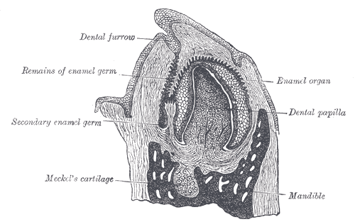 Vertical section of the mandible of an early human fetus. X 25.

External links
Template:Gray's Template:Embryology of head and neck Template:WH Template:WikiDoc Sources