Lumbar plexus

|
WikiDoc Resources for Lumbar plexus |
|
Articles |
|---|
|
Most recent articles on Lumbar plexus Most cited articles on Lumbar plexus |
|
Media |
|
Powerpoint slides on Lumbar plexus |
|
Evidence Based Medicine |
|
Clinical Trials |
|
Ongoing Trials on Lumbar plexus at Clinical Trials.gov Trial results on Lumbar plexus Clinical Trials on Lumbar plexus at Google
|
|
Guidelines / Policies / Govt |
|
US National Guidelines Clearinghouse on Lumbar plexus NICE Guidance on Lumbar plexus
|
|
Books |
|
News |
|
Commentary |
|
Definitions |
|
Patient Resources / Community |
|
Patient resources on Lumbar plexus Discussion groups on Lumbar plexus Patient Handouts on Lumbar plexus Directions to Hospitals Treating Lumbar plexus Risk calculators and risk factors for Lumbar plexus
|
|
Healthcare Provider Resources |
|
Causes & Risk Factors for Lumbar plexus |
|
Continuing Medical Education (CME) |
|
International |
|
|
|
Business |
|
Experimental / Informatics |
Overview
The lumbar plexus is a nervous plexus in the lumbar region of the body. It is formed by the loops of communication between the anterior divisions of the first three and the greater part of the fourth lumbar nerves; the first lumbar often receives a branch from the last thoracic nerve.
It is situated in the posterior part of the Psoas major, in front of the transverse processes of the lumbar vertebræ.
The mode in which the plexus is arranged varies in different subjects.
Branches
The lumbar plexus differs from the brachial plexus in not forming an intricate interlacement, but the several nerves of distribution arise from one or more of the spinal nerves, in the following manner: the first lumbar nerve, frequently supplemented by a twig from the last thoracic, splits into an upper and lower branch; the upper and larger branch divides into the iliohypogastric and ilioinguinal nerves; the lower and smaller branch unites with a branch of the second lumbar to form the genitofemoral nerve.
The remainder of the second lumbar nerve, and the third and fourth lumbar nerves, divide into ventral and dorsal divisions.
The ventral division of the second lumbar nerve unites with the ventral divisions of the third and fourth lumbar nerves to form the obturator nerve.
The dorsal divisions of the second and third nerves divide into two branches, a smaller branch from each uniting to form the lateral femoral cutaneous nerve, and a larger branch from each joining with the dorsal division of the fourth nerve to form the femoral nerve.
The accessory obturator, when it exists, is formed by the union of two small branches given off from the third and fourth nerves.
Mnemonic
One mnemonic used to remember this is "Interested in getting laid (or lunch) on Friday, Larry?"
| Division | Name | Source | Target |
| Main | Iliohypogastric nerve | 1 L. | Skin over the lateral gluteal region and above the pubis [1] |
| Main | Ilioinguinal nerve | 1 L. | Skin over the root of the penis and upper part of the scrotum (male), skin covering the mons pubis and labium majus (female) |
| Main | Genitofemoral nerve | 1, 2 L. | Genital Branch: Cremaster muscle, skin of scrotum/labia majora Femoral Branch: Skin on anterior thigh |
| Dorsal | Lateral femoral cutaneous | 2, 3 L. | Skin on the lateral part of the thigh |
| Ventral | Obturator nerve (and Accessory obturator nerve, when present) | 2, 3, 4 L. | Medial compartment of thigh |
| Dorsal | Femoral nerve | 2, 3, 4 L. | Anterior compartment of thigh |
| Ventral | Lumbosacral trunk | 4, 5L., 1, 2, 3, 4 S. | Sacral plexus |
Additional images
-
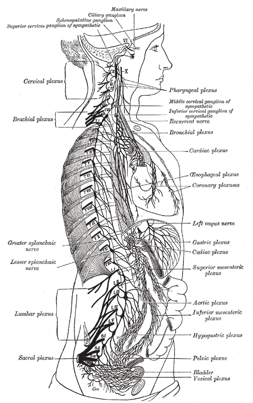 The right sympathetic chain and its connections with the thoracic, abdominal, and pelvic plexuses.

References
External links
Template:Gray's Template:Anatomy-stub Template:Spinal nerves Template:Lumbosacral plexus