Sacral plexus
Template:Infobox Nerve Editor-In-Chief: C. Michael Gibson, M.S., M.D. [1]
Overview
In human anatomy, the sacral plexus is a nerve plexus emerging from the sacral vertebrae (S1-S4), and which provides nerves for the pelvis and lower limbs.
Structure
The sacral plexus is formed by::
- the lumbosacral trunk
- the anterior division of the first sacral nerve
- portions of the anterior divisions of the second and third sacral nerves
The nerves forming the sacral plexus converge toward the lower part of the greater sciatic foramen, and unite to form a flattened band, from the anterior and posterior surfaces of which several branches arise.
The band itself is continued as the sciatic nerve, which splits on the back of the thigh into the tibial nerve and common fibular nerve; these two nerves sometimes arise separately from the plexus, and in all cases their independence can be shown by dissection.
Often, the sacral plexus and the lumbar plexus are considered to be one large nerve plexus, the lumbosacral plexus. The lumbosacral trunk connects the two plexuses.
Relations
The sacral plexus lies on the back of the pelvis between the piriformis muscle and the pelvic fascia. In front of it are the internal iliac artery, internal iliac vein, the ureter, and the sigmoid colon. The superior gluteal artery and vein run between the lumbosacral trunk and the first sacral nerve, and the inferior gluteal artery and vein between the second and third sacral nerves.
All the nerves entering the plexus, with the exception of the third sacral, split into ventral and dorsal divisions, and the nerves arising from these are as follows:
| Nerve | Segments | Muscles | Cutaneous |
| Nerve to quadratus femoris | L4-S1 | gemellus inferior, quadratus femoris | |
| Superior gluteal nerve | L4-S1 | gluteus medius, gluteus minimus, tensor fasciae latae | |
| Sciatic nerve | L4-S3 | ||
| * Tibial nerve | L4-S3 | posterior compartment | posterolateral leg and foot - medial sural cutaneous nerve |
| * Common fibular nerve | L4-S3 | anterior and lateral compartments | anterolateral leg and foot - Lateral sural cutaneous nerve, medial dorsal cutaneous nerve, intermediate dorsal cutaneous nerve |
| Nerve to obturator internus | L5-S2 | gemellus superior, obturator internus | |
| Inferior gluteal nerve | L5-S2 | gluteus maximus | |
| Nerve to piriformis | S1-S2 | piriformis | |
| Posterior cutaneous nerve of thigh | S1-S3 | - | thigh |
| Pudendal nerve | S2-S4 | bulbospongiosus, deep transverse perineal, ischiocavernosus, sphincter urethrae, superficial transverse perineal | clitoris, penis |
| Coccygeal nerve | S4-Co1 | - | perineum |
Additional images
-
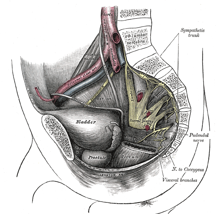
Dissection of side wall of pelvis showing sacral and pudendal plexuses.
-
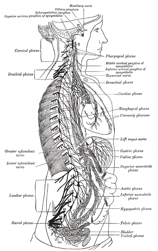
The right sympathetic chain and its connections with the thoracic, abdominal, and pelvic plexuses.


See also
External links
- Template:GPnotebook
- Lumbosacral+Plexus at the US National Library of Medicine Medical Subject Headings (MeSH)
- Template:ViennaCrossSection
- Template:MedicalMnemonics
- Template:EMedicineDictionary
- Illustration at preventdisease.com
- Illustration at backpain-guide.com
Template:Gray's
Template:Spinal nerves
Template:Lumbosacral plexus