Globus pallidus
The globus pallidus (Latin for "pale globe") is a sub-cortical structure of the brain. It is a major element of the basal ganglia system. In this system, it is a major element of the basal ganglia core; made up of the striatum and its direct targets: globus pallidus and substantia nigra. The last two are made up of the same neuronal elements, have a similar main afferent, the dorsal striatum, a similar synaptology and do not receive cortical afferents.
History of name
The origin of the name is not established. It was known by Dejerine (1906) but not by Santiago Ramón y Cajal (1909-1911). As the elements in no way have the shape of a globe, Foix and Nicolesco (1925), the Vogts (1941), Crosby et all.(1962) followed by the Terminologia anatomica proposed the simpler term (neutral adjective) of pallidum (pale). During a long period the globus pallidus was unduly linked to the putamen in the lentiform nucleus (nucleus lenticularis or lentiformis). This was a heterogeneous anatomical entity that is part of the striatum rather than the pallidum. The link with the substantia reticulata was stressed very early on due to the similarities in dendritic arborisation, but in spite of solid arguments this link is still not widely accepted. The two however constitute a particular set of the basal ganglia system (the pallidonigral set).
Parts
In primates, the dorsal pallidum, or globus pallidus, is divided into two segments by the medial medullary lamina. A frequent nomenclature uses the adjectives internal and external to refer to the two divisions of the globus pallidus. The medial segment of the dorsal pallidum, internal globus pallidus (GPi) and lateral division of the dorsal pallidum, external globus pallidus (GPe) are thus the two parts of the dorsal pallidum that are two closed nuclei surrounded everywhere by myelinic walls.
The ventral pallidum lies within the substantia innominata (Latin for un-named substance) and receives efferent connections from the ventral striatum (nucleus accumbens and olfactory tubercle). The ventral pallidum projects to the dorsomedial nucleus of the dorsal thalamus, which projects to the prefrontal cortex. The ventral pallidum also projects to the pedunclopontine nucleus and tegmental motor area. The function of the ventral pallidum is limbic-somatic motor interface for the planning and inhibition of movements from the dorsal striatopallidal complex.
Structure
Pallidal nuclei are made up of the same neuronal components. In primates, almost all pallidal neurons are very large, parvalbumin positive, with very large dendritic arborizations. These have the peculiarity of having the three dimensional shape of flat discs, parallel one to the other, parallel to the border of the pallidum[1] and perpendicular to the afferent striatopallidal axons.[2] There are only a few small local circuitry neurons.
The globus pallidus is traversed by the numerous myelinated axons of the striato-pallidonigral bundle that give it the pale appearance from which it is named.
The ultrastructure is very peculiar as the long dendrites are everywhere, without discontinuity, covered by synapses.[3][4]
Pallidonigral pacemaker
The two pallidal nuclei and the two nigral (pars compacta and pars reticulata) parts constitute a high frequency autonomous pacemaker[5] (see primate basal ganglia system)
Common afferences
The two parts receive successively a large quantity of GABAergic axonal terminal arborisations from the striatum through the dense striato-pallidonigral bundle. The synaptology is very peculiar (see primate basal ganglia system).[3][4]The striatal afference contribute for more than 90% of synapses. The two pallidal nuclei receives dopaminergic axons from the pars compacta of the substantia nigra.
Other connections and subsystems

ROSTRAL: striatum, globus pallidus (GPe and GPi)
CAUDAL: subthalamic nucleus (STN), substantia nigra (SN)
External Globus Pallidus (GPe)
The internal globus pallidus receives a strong glutamatergic projection from the subthalamic nucleus. The two form a particular system: a coupled pacemaker.
The axons of the external globus pallidum go essentially to the subthalamic nucleus. They go also to other elements of the basal ganglia system, the striatum, the substantia nigra pars reticulata and the internal globus pallidus, where they release the neurotransmitter GABA. GPe is particular in comparison to the other elements of the set by the fact that it does not work as an output base of the basal ganglia (not sending axons to the thalamus) but as the main regulator of the basal ganglia system. It is sometimes used as a target for deep brain stimulation as a treatment for Parkinson's disease.
Internal Globus Pallidus (GPi)
The internal segment of the globus pallidus, GPi is one of the output nuclei of the basal ganglia (the other being the substantia nigra pars reticulata). The GABA-containing neurons send their axons to specific nuclei of the dorsal thalamus (VA and VL), to the centremedian complex and to the pedunculopontine complex.[6][7]
The efferent bundle is constituted first of the ansa and fasciculus lenticularis, then crosses the internal capsule as the Edinger's comb system then arrives at the laterosuperior corner of the subthalamic nucleus and constitutes the Forel's field H2, then H and suddenly changes its direction to form H1 that goes to the inferior part of the thalamus. The distribution of axonal islands is widespread in the lateral region of the thalamus. The innervation of the central region is done by collaterals.[8]
References
- ↑ Yelnik, J., Percheron, G., and François, C. (1984) A Golgi analysis of the primate globus pallidus. II- Quantitative morphology and spatial orientation of dendritic arborisations. J. Comp. Neurol. 227:200-213
- ↑ Percheron, G.,Yelnik, J. and François. C. (1984) A Golgi analysis of the primate globus pallidus. III-Spatial organization of the striato-pallidal complex. J. Comp. Neurol. 227: 214-227
- ↑ 3.0 3.1 Fox, C.A., Andrade, A.N. Du Qui, I.J., Rafols, J.A. (1974) The primate globus pallidus. A Golgi and electron microscopic study. J. Hirnforsch. 15: 75-93
- ↑ 4.0 4.1 di Figlia, M., Pasik, P., Pasik, T. (1982) A Golgi and ultrastructural study of the monkey globus pallidus. J. Comp. Neurol. 212: 53-75
- ↑ Surmeier, D.J., Mercer, J.N. and Savio Chan, C. (2005) Autonomous pacemakers in the basal ganglia: who needs excitatory synapses anyway? Cur. Opin.Neurobiol. 15:312-318.
- ↑ Nauta, W.J.H. and Mehler, W.R. (1966) Projections of the lentiform nucleus in the monkey. Brain Res. 1: 3-42
- ↑ Percheron, G., François, C., Talbi, B., Yelnik, J., Fenelon, G. (1996) The primate motor thalamus. Brain Res. Rev. 22: 93-181
- ↑ Arrechi-Bouchhioua, P., Yelnik, K., François, C..,Percheron. G., Tandé, D. (1997) Three-dimensional morphology and distribution of pallidal axons projecting to both the lateral region of the thalamus and the central complex in primates. Brain Res. 754: 311-314
Additional images
-
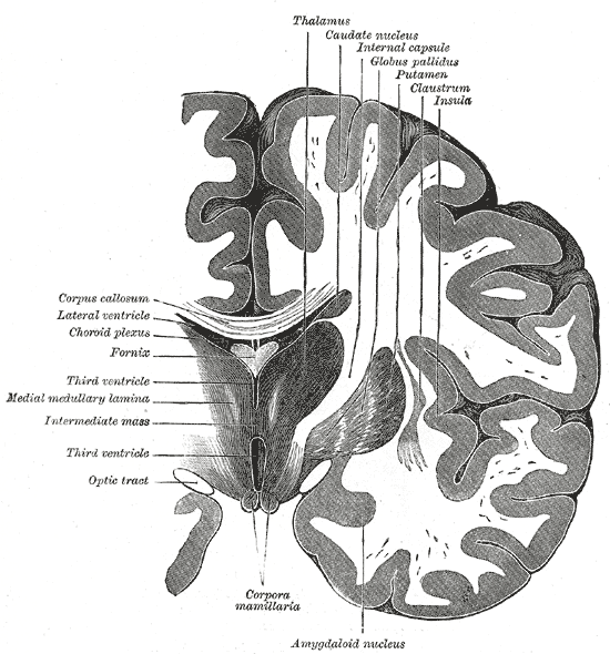 Coronal section of brain through intermediate mass of third ventricle.
 -
 Coronal section of brain through anterior commissure.
 -
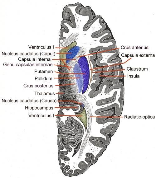 Horizontal section of right cerebral hemisphere.
 -
Connectivity Diagram showing glutamatergic pathways as red, dopaminergic as magenta and GABA pathways as blue.


