External jugular vein
Template:Infobox Vein Editor-In-Chief: C. Michael Gibson, M.S., M.D. [1]
The external jugular vein receives the greater part of the blood from the exterior of the cranium and the deep parts of the face, being formed by the junction of the posterior division of the posterior facial with the posterior auricular vein.
It commences in the substance of the parotid gland, on a level with the angle of the mandible, and runs perpendicularly down the neck, in the direction of a line drawn from the angle of the mandible to the middle of the clavicle at the posterior border of the Sternocleidomastoideus.
In its course it crosses the Sternocleidomastoideus obliquely (does it cross it anteriorly?), and in the subclavian triangle perforates the deep fascia, and ends in the subclavian vein(does it ever end in the brachiocephalic trunk on the right?), lateral to or in front of the Scalenus anterior.
It is separated from the Sternocleidomastoideus by the investing layer of the deep cervical fascia, and is covered by the Platysma, the superficial fascia, and the integument; it crosses the cutaneous cervical nerve, and its upper half runs parallel with the great auricular nerve.
The external jugular vein varies in size, bearing an inverse proportion to the other veins of the neck, it is occasionally double.
It is provided with two pairs of valves, the lower pair being placed at its entrance into the subclavian vein, the upper in most cases about 4 cm. above the clavicle. The portion of vein between the two sets of valves is often dilated, and is termed the sinus.
These valves do not prevent the regurgitation of the blood, or the passage of injection from below upward.
Tributaries
This vein receives the occipital occasionally, the posterior external jugular, and, near its termination, the transverse cervical, transverse scapular, and anterior jugular veins; in the substance of the parotid, a large branch of communication from the internal jugular joins it.
Additional images
-
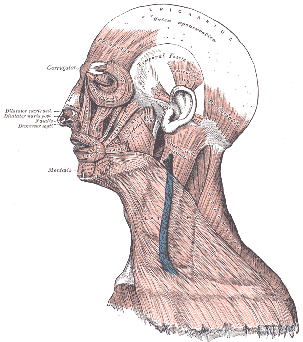 Muscles of the head, face, and neck.
 -
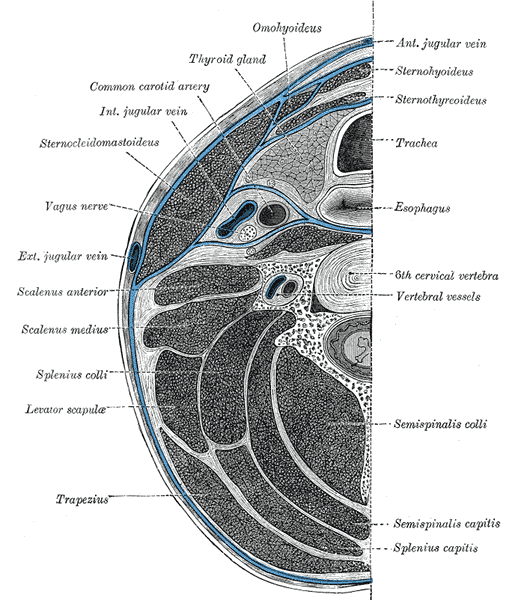 Section of the neck at about the level of the sixth cervical vertebra.
 -
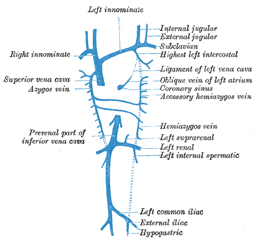 Diagram showing completion of development of the parietal veins.
 -
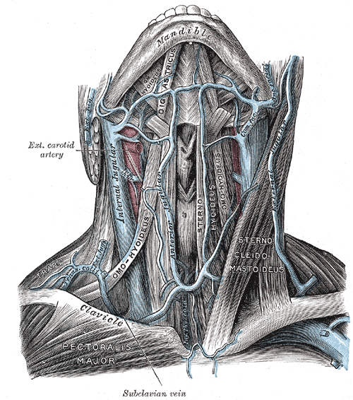 The veins of the neck, viewed from the front.
 -
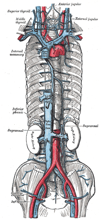 The venæ cavæ and azygos veins, with their tributaries.
 -
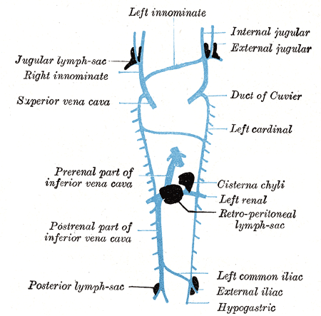 Scheme showing relative positions of primary lymph sacs.


