Echinococcosis pathophysiology
|
Echinococcosis Microchapters |
|
Diagnosis |
|---|
|
Treatment |
|
Case Studies |
|
Echinococcosis pathophysiology On the Web |
|
American Roentgen Ray Society Images of Echinococcosis pathophysiology |
|
Risk calculators and risk factors for Echinococcosis pathophysiology |
Editor-In-Chief: C. Michael Gibson, M.S., M.D. [1] Associate Editor-In-Chief: Mahshid Mir, M.D. [2] Cafer Zorkun, M.D., Ph.D. [3]; Kalsang Dolma, M.B.B.S.[4]
Overview
The transmission of echinococcosis from the definitive host to the intermediate host is by ingestion of embryonated eggs passed in the feces. Once the eggs are ingested they hatch in the small intestine and develop into onchospheres. There oncospheres reach various organs by migration through the vascular system and develop into cysts producing protoscolices. The definitive host is infected by ingestion of the organs infected with the cysts. After ingestion of the cysts they evaginate and invade the intestinal mucosa and develop into adult worms.
Pathophysiology
Life cycle

(1)The adult Echinococcus granulosus (2) Embryonated eggs (3) Oncosphere (4) Cyst (5) Protoscolices (6) Protoscolices evaginating
Trasmission of infection[1][2][3][4]
- The transmission of echinococcosis from the definitive host (dogs and other carnivores) to the intermediate host (sheep, goats, swine etc) occurs by the ingestion of embryonated eggs passed in the feces.
- The definitive host is infected by the ingestion of cyst containing organs of the infected intermediate host (sheep, goats, swine, etc).
Pathogenesis[1][2][3][4]
- The embronated eggs are excreted in the feces of the definitive host, which include dogs and other carnivores.
- The intermediate hosts include: sheep, cattle, horses and camel. Once ingested the eggs hatch in the small bowel and release oncospheres.
- The onchospheres penetrate the intestinal wall and migrate through the vascular system to organs such as liver and lung.
- In the lungs and liver the oncospheres develop into a cyst producing protoscolices and daughter cysts which fill the interior of the cyst.
- The definite host will be infected if they ingest organs containing cysts.
- Once ingested, the protoscolices evaginate and attach the intestinal mucosa.
- In 32 to 80 days after evagination the protoscolices develop into adult tapeworm.
- The life cycle of E. multilocularis is similar to the life cycle of Echinococcus granulosus, but with the following differences: The definitive hosts are foxes, and to a lesser extent dogs, cats, coyotes and wolves. The intermediate host are small rodents and the larval growth (in the liver) remains indefinitely in the proliferative stage, resulting in invasion of the surrounding tissues.
- In the life cycle of E. vogeli the definitive hosts are bush dogs and dogs. The intermediate hosts are rodents and the larval stage in the liver, lung develops both externally and internally, resulting in multiple vesicles.
- E. oligarthrus (up to 2.9 mm long) has a life cycle that involves wild felids as definitive hosts and rodents as intermediate hosts. Humans become infected by ingesting eggs , with resulting release of oncospheres in the intestine and the development of cysts in various organs.
Gross Pathology[4]
The most important gross pathological features of echinococcus cysts are:
- Cysts of E. granulosis (cystic hydatid disease):
- Cysts tend to be:
- Filled with clear fluid
- White appearance
- Solitary
- Unilocular
- Mostly involve right lobe of liver
- Viable cysts are filled with a colorless fluid that contains daughter cysts and brood capsules with scolices
- Daughter cysts may also be present outside the fibrous layer of the cyst, that are called as extracapsular or satellite cysts
- Cysts tend to be:
- Cysts of E. multilocularis (alveolar hydatid disease):
- Numerous small and irregular cysts
- Mostly smaller than 2 cm
- Appears infiltrative


Microscopic pathology[4]
On microscopic pathology of the tissues, the most important findings include:
- Cysts can be found in any part of the body, but are most common in the liver, lung and central nervous system
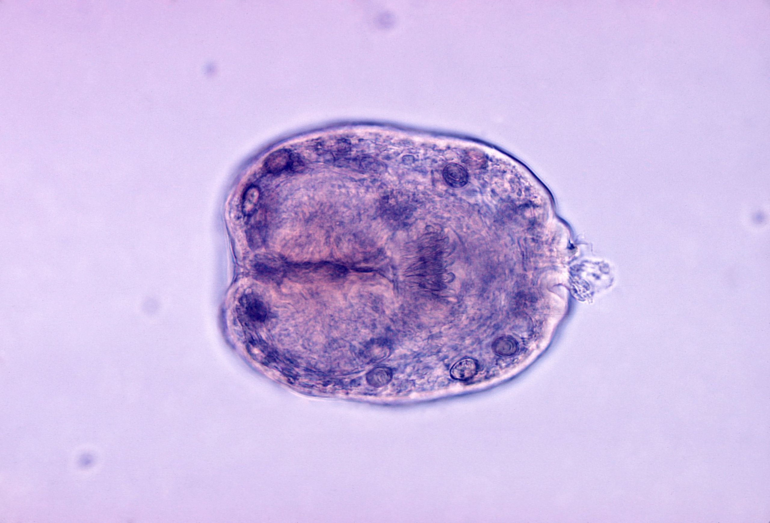
- Echinococcus granulosus cyst:
- Cyst wall composed of an acellular laminated external layer and a thin, germinal (nucleated) inner layer
- Brood capsule with protoscoleces inside


Echinococcus granulosus scolex close-up
References
- ↑ 1.0 1.1 Rasheed K, Zargar SA, Telwani AA (2013). "Hydatid cyst of spleen: a diagnostic challenge". N Am J Med Sci. 5 (1): 10–20. doi:10.4103/1947-2714.106184. PMC 3560132. PMID 23378949.
- ↑ 2.0 2.1 Pakala T, Molina M, Wu GY (2016). "Hepatic Echinococcal Cysts: A Review". J Clin Transl Hepatol. 4 (1): 39–46. doi:10.14218/JCTH.2015.00036. PMC 4807142. PMID 27047771.
- ↑ 3.0 3.1 Siracusano A, Delunardo F, Teggi A, Ortona E (2012). "Host-parasite relationship in cystic echinococcosis: an evolving story". Clin. Dev. Immunol. 2012: 639362. doi:10.1155/2012/639362. PMC 3206507. PMID 22110535.
- ↑ 4.0 4.1 4.2 4.3 "CDC - DPDx - Echinococcosis".