Bronchus
|
WikiDoc Resources for Bronchus |
|
Articles |
|---|
|
Most recent articles on Bronchus |
|
Media |
|
Evidence Based Medicine |
|
Clinical Trials |
|
Ongoing Trials on Bronchus at Clinical Trials.gov Clinical Trials on Bronchus at Google
|
|
Guidelines / Policies / Govt |
|
US National Guidelines Clearinghouse on Bronchus
|
|
Books |
|
News |
|
Commentary |
|
Definitions |
|
Patient Resources / Community |
|
Directions to Hospitals Treating Bronchus Risk calculators and risk factors for Bronchus
|
|
Healthcare Provider Resources |
|
Causes & Risk Factors for Bronchus |
|
Continuing Medical Education (CME) |
|
International |
|
|
|
Business |
|
Experimental / Informatics |
Editor-In-Chief: C. Michael Gibson, M.S., M.D. [1]
Overview
A bronchus (plural bronchi, adjective bronchial) is a caliber of airway in the respiratory tract that conducts air into the lungs. No gas exchange takes place in this part of the lungs.
Anatomy
The trachea (windpipe) divides into two main bronchi (also mainstem bronchi), the left and the right, at the level of the sternal angle. The right main bronchus is wider, shorter, and more vertical than the left main bronchus. The right main bronchus subdivides into three segmental bronchi while the left main bronchus divides into two. The lobar bronchi divide into tertiary bronchi. Each of the segmental bronchi supplies a bronchopulmonary segment. A bronchopulmonary segment is a division of a lung that is separated from the rest of the lung by a connective tissue septum. This property allows a bronchopulmonary segment to be surgically removed without affecting other segments. There are ten segments per lung, but due to anatomic development, several segmental bronchi in the left lung fuse, giving rise to eight. The segmental bronchi divide into many primary bronchioles which divide into terminal bronchioles, each of which then gives rise to several respiratory bronchioles, which go on to divide into 2 to 11 alveolar ducts. There are 5 or 6 alveolar sacs associated with each alveolar duct. The alveolus is the basic anatomical unit of gas exchange in the lung.
There is hyaline cartilage present in the bronchi, present as irregular rings in the larger bronchi (and not as regular as in the trachea), and as small plates and islands in the smaller bronchi. Smooth muscle is present continuously around the bronchi.
In the mediastinum, at the level of the fifth thoracic vertebra, the trachea divides into the right and left primary bronchi. The bronchi branch into smaller and smaller passageways until they terminate in tiny air sacs called alveoli.
The cartilage and mucous membrane of the primary bronchi are similar to that in the trachea. As the branching continues through the bronchial tree, the amount of hyaline cartilage in the walls decreases until it is absent in the smallest bronchioles. As the cartilage decreases, the amount of smooth muscle increases. The mucous membrane also undergoes a transition from ciliated pseudostratified columnar epithelium to simple cuboidal epithelium to simple squamous epithelium.
The alveolar ducts and alveoli consist primarily of simple squamous epithelium, which permits rapid diffusion of oxygen and carbon dioxide. Exchange of gases between the air in the lungs and the blood in the capillaries occurs across the walls of the alveolar ducts and alveoli.
Role in disease
Bronchitis is defined as inflammation of the bronchi. There are two main types: acute and chronic. Acute bronchitis is usually caused by viral or bacterial infections. Chronic bronchitis is a form of COPD, usually associated with smoking or long-term exposure to irritants.
Asthma is hyperreactivity of the bronchi with an inflammatory component, often in response to allergens.
While the left mainstem bronchus departs from the trachea at an angle, the right mainstem bronchus is almost a vertical continuation of the trachea. This anatomy predisposes the right lung to several problems:
- If food, liquids, or foreign bodies are aspirated, they often will lodge in the right mainstem bronchus. Aspiration pneumonia may result.
- If the endotracheal tube used for intubation is inserted too far, it usually lodges in the right mainstem bronchus. This allows ventilation of the right lung, but leaves the left lung useless.
- Patients with inadequate cough reflexes may develop chronic right middle lobe lung infections such as the Lady Windermere Syndrome.
Additional images
-
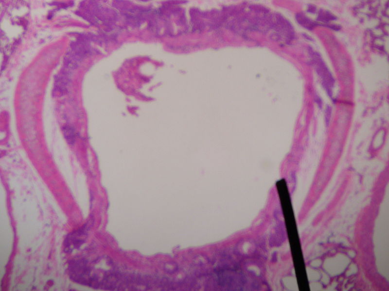 Cross sectional cut of a human secondary bronchus
 -
 Transverse section of thorax, showing relations of pulmonary artery.


References
- Moore, Keith L. and Arthur F. Dalley. Clinically Oriented Anatomy, 4th ed. (1999). ISBN 0-7817-5936-6
ca:Bronqui da:Bronkie de:Bronchialsystem dv:ވައި ނޮޅީގެ ކަފި eo:Bronko id:Bronkus it:Bronco he:סמפון la:Bronchus lt:Bronchas nl:Luchtpijpvertakking no:Bronkietreet fi:Keuhkoputki sv:Bronk uk:Бронхи