Respiratory bronchiole
Jump to navigation
Jump to search
The respiratory bronchioles are the beginning of the respiratory segment of the airway and are just distal to the terminal bronchioles (which are the last segment of the conducting airway). The epithelium in this segment is simple cuboidal. The respiratory bronchioles are interrupted by alveoli which are thin walled evaginations. Alveolar ducts are distal continuations of the respiratory bronchioles.
Additional images
-
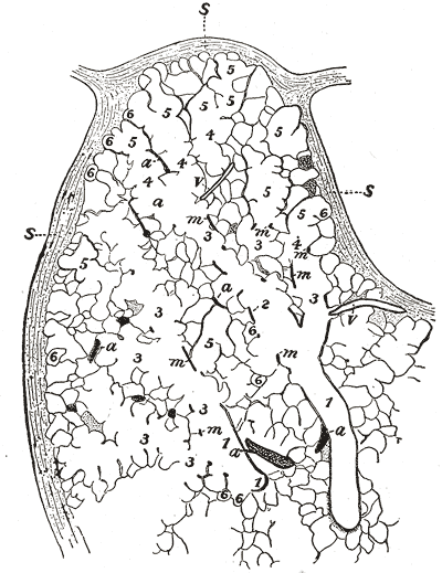 Part of a secondary lobule from the depth of a human lung, showing parts of several primary lobules.

External links
- Diagram at davidson.edu
- Template:EMedicineDictionary
- Histology image: 13606loa – Histology Learning System at Boston University
- Histology at umdnj.edu