Bacterial pneumonia pathophysiology

|
Bacterial pneumonia Microchapters |
|
Diagnosis |
|
Treatment |
|
Case Studies |
|
Bacterial pneumonia pathophysiology On the Web |
|
American Roentgen Ray Society Images of Bacterial pneumonia pathophysiology |
|
Risk calculators and risk factors for Bacterial pneumonia pathophysiology |
Editor(s)-in-Chief: C. Michael Gibson, M.S., M.D. [1] Phone:617-632-7753; Philip Marcus, M.D., M.P.H.[2] Associate Editor(s)-in-Chief: Arooj Naz, M.B.B.S
Overview
Causative bacteria can be inhaled from the surrounding environment but organisms are also commonly found within the upper respiratory tract of many individuals from where they can be directed towards the alveoli. Once in the bloodstream of the lungs, the immune system triggers a response by mobilizing white blood cells into the affected area. Neutrophils and cytokine help in ridding the body of infectious organisms but also result in systemic symptoms such as fever, chills, and fatigue. If bacteria enters the systemic blood stream, sepsis may ensue and can involve other organs including the brain, kidney, and heart.
Pathophysiology
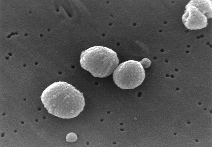
Bacteria[1]
- Bacteria and fungi also typically enter the lung with inhalation, though they can reach the lung through the bloodstream if other parts of the body are infected. Most commonly, bacteria enter via aspiration, but infection via hematogenous spread may also occur.[2]
- Often, bacteria live in parts of the upper respiratory tract and are constantly being inhaled into the alveoli.
- Once inside the alveoli, bacteria and fungi travel into the spaces between the cells and also between adjacent alveoli through connecting pores.
- This invasion triggers the immune system to respond by sending white blood cells responsible for attacking microorganisms (neutrophils) to the lungs. The neutrophils engulf and kill the offending organisms but also release cytokines which result in a general activation of the immune system.
- Fever, chills, and fatigue are common in CAP. The neutrophils, bacteria, and fluid leaked from surrounding blood vessels fill the alveoli and result in impaired oxygen transportation.
- Bacteria often travel from the lung into the blood stream and can result in serious illness such as septic shock. Inflammation and further damage is further exacerbated by the release of cytokines such as IL-1 and TNF leading to multiorgan failure and septic shock.
- Septic shock results in low blood pressure leading to damage in multiple parts of the body including the brain, kidney, and heart.