Babesiosis pathophysiology
|
Babesiosis Microchapters |
|
Diagnosis |
|---|
|
Treatment |
|
Case Studies |
|
Babesiosis pathophysiology On the Web |
|
American Roentgen Ray Society Images of Babesiosis pathophysiology |
|
Risk calculators and risk factors for Babesiosis pathophysiology |
Editor-In-Chief: C. Michael Gibson, M.S., M.D. [1]; Associate Editor(s)-in-Chief: Tamar Sifri [2] Associate Editor(s)-in-Chief: Ilan Dock, B.S.
Overview
Babesia parasites reproduce in red blood cells, where they can be seen as cross-shaped inclusions (4 merozoites asexually budding but attached together forming a structure looking like a "Maltese Cross") and cause hemolytic anemia, quite similar to malaria. Note that unlike the Plasmodium parasites that cause malaria, Babesia species lack an exo-erythrotic phase, so the liver is usually not affected. The cycle depends on two primary components, an infected vertebrate host and an infected tick. Transmission from a tick to a human occurs over the course of a complete blood meal. Inoculation rates will rapidly increase in direct correlation with the span of the tick's blood meal.
Life Cycle

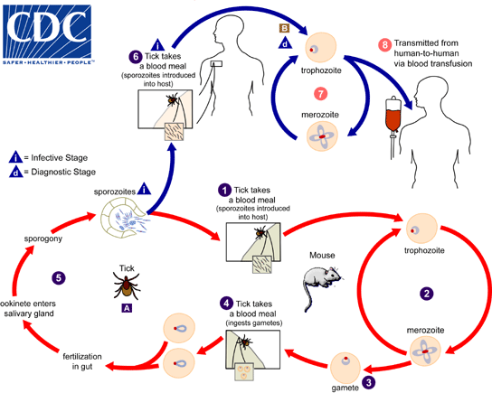
- The Babesia microti life cycle involves two primary components, an infected vertebrate host (primarily the white-footed mouse "Peromyscus leucopus"), and a tick in the genus Ixodes. [1]
Pathogenic events within the tick
Tick Ingestion of Babesia spp.
- Babesia organisms are detectable within 10 hours of tick ingestion.
- Parasitic organisms may still be detectable within erythrocytes until this point, however gametocytes begin to develop new organelles within 46 to 60 hours of ingestion.
- A notable physiological event occurs as gametocytes develop an arrowhead-shaped organelle.
- Research suggests that these arrowhead like structures are involved in gamete fusion.[2]
Zygote Development
- Zygotes maintain gamete's arrowhead like structures.
- These structures are used by the Zygote to penetrate epithelial cells within the tick's gut. Occurring within 80h of ingestion.
- Parasitic Organisms travel from epithelial cells in the tick gut to the salivary acini by means of the hemolymph.
Sporozoite Development
- Three notable stages of sporozoite development occur within the tick's salivary glands.
- Formation of a multinucleate sporoblast within a hypertrophied host cell. (Results in three dimensional branching and an eventual foundation for sporozoite budding.)
- Host begins feeding, resulting in the formation of sporozoite's specialized organelles.
- Budding of sporozoites. A single sporoblast may result in 5,000 to 10,000 sporozoites.[2]
Transmission
Tick to Mouse Transmission
- During a blood meal, a Babesia-infected tick introduces sporozoites into the mouse host.
- Sporozoites enter erythrocytes and undergo asexual reproduction (budding).
- In the blood, some parasites differentiate into male and female gametes, although these cannot be distinguished by light microscopy.
- The definitive host is the tick. [2]
Transmission to the Human Host
- Transmission occurs during a blood meal.
- Parasitic organism are transmitted through in the tick's saliva.
- Tick transmission is an efficient transmission method since the tick's saliva contains anti-inflammatory and immunosuppressive properties.
- Enabling transmission to occur largely undetected.
Transovarial transmission (also known as vertical, or hereditary, transmission) has been documented for "large" Babesia spp. but not for the "small" babesiae, such as B. microti (A).
Pathogenesis Within the Vertebrate Host
- Several thousand sporozoites are transmitted in the dermis surrounding the tick's mouth.
- A successful inoculation requires 10,000-25,000 sporozoites. Therefore there is a direct correlation between the duration of tick feeding and probability of contracting the disease.
- Infection rates reach nearly 100% when an infected tick feed to repletion.[2]
- Sporozoites primarily infect erythrocytes.
- Certain species of Babesia will infect lymphocytes.
- Infection results in multinucleate schizont which bud into merozoites and cause cell lysing.
- Merozoites infect further erythrocytes through the formation of a parsitophorous vacuole, a process called invagination.
- Post-invasion the vacuole membrane disintegrates revealing a remaining single membrane, unlike plasmodium infection which retains both the host and parasitic membrane.[2]
References
- ↑ CDC Babesiosis. Biology. http://www.cdc.gov/parasites/babesiosis/biology.html Accessed December 21, 2016
- ↑ 2.0 2.1 2.2 2.3 2.4 Homer, Mary J.; Aguilar-Delfin, Irma; Telford III, Sam R.; Krause ,Peter J.; Persing, David H.; Babesiosis. Clinical Microbiology Reviews. 2000;13(3):451. http://cmr.asm.org/content/13/3/451.full Accessed on January 14, 2016.