Aortic arches
|
WikiDoc Resources for Aortic arches |
|
Articles |
|---|
|
Most recent articles on Aortic arches Most cited articles on Aortic arches |
|
Media |
|
Powerpoint slides on Aortic arches |
|
Evidence Based Medicine |
|
Clinical Trials |
|
Ongoing Trials on Aortic arches at Clinical Trials.gov Trial results on Aortic arches Clinical Trials on Aortic arches at Google
|
|
Guidelines / Policies / Govt |
|
US National Guidelines Clearinghouse on Aortic arches NICE Guidance on Aortic arches
|
|
Books |
|
News |
|
Commentary |
|
Definitions |
|
Patient Resources / Community |
|
Patient resources on Aortic arches Discussion groups on Aortic arches Patient Handouts on Aortic arches Directions to Hospitals Treating Aortic arches Risk calculators and risk factors for Aortic arches
|
|
Healthcare Provider Resources |
|
Causes & Risk Factors for Aortic arches |
|
Continuing Medical Education (CME) |
|
International |
|
|
|
Business |
|
Experimental / Informatics |
Editor-In-Chief: C. Michael Gibson, M.S., M.D. [1]
- This article focuses upon the multiple aortic arches present in the embryo. For the single structure present in the adult, see Aortic arch
Overview
The aortic arches are a series of six paired embryological vascular structures which give rise to several major arteries. They are ventral to the dorsal aorta.
Specific arches
Arches 1 and 2
The first and second arches disappear early, but the dorsal end of the second gives origin to the stapedial artery, a vessel which atrophies in humans but persists in some mammals. It passes through the ring of the stapes and divides into supraorbital, infraorbital, and mandibular branches which follow the three divisions of the trigeminal nerve. The infraorbital and mandibular arise from a common stem, the terminal part of which anastomoses with the external carotid.
On the obliteration of the stapedial artery this anastomosis enlarges and forms the internal maxillary artery, and the branches of the stapedial artery are now branches of this vessel.
The common stem of the infraorbital and mandibular branches passes between the two roots of the auriculotemporal nerve and becomes the middle meningeal artery; the original supraorbital branch of the stapedial is represented by the orbital twigs of the middle meningeal.
Arch 3
The third aortic arch constitutes the commencement of the internal carotid artery, and is therefore named the carotid arch.
Arch 4
The fourth right arch forms the right subclavian as far as the origin of its internal mammary branch; while the fourth left arch constitutes the arch of the aorta between the origin of the left carotid artery and the termination of the ductus arteriosus.
Arch 5
The fifth arch disappears on both sides.
Arch 6
The sixth right arch disappears; the sixth left arch gives off the pulmonary arteries and forms the ductus arteriosus; this duct remains pervious during the whole of fetal life, but is obliterated a few days after birth.
His showed that in the early embryo the right and left arches each gives a branch to the lungs, but that later both pulmonary arteries take origin from the left arch.
Additional images
-
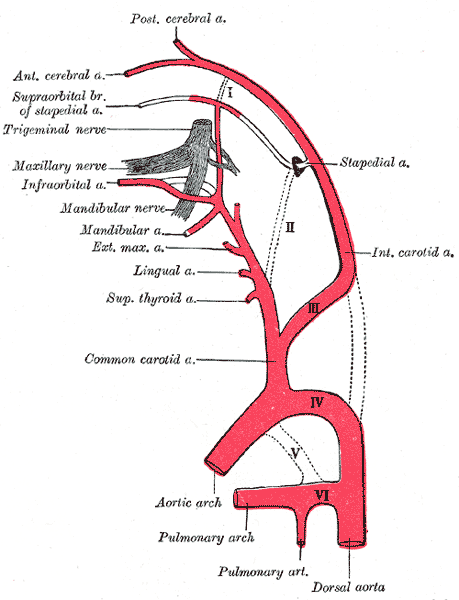 Diagram showing the origins of the main branches of the carotid arteries.

External links
- Template:EmbryologyTemple
- Diagram at University of Michigan
- Template:EmbryologyUNC
See Also
Template:Gray's Template:Development of circulatory system