Ventricular septal defect: Difference between revisions
No edit summary |
No edit summary |
||
| Line 141: | Line 141: | ||
==[[Ventricular septal defect overview|Overview]]== | ==[[Ventricular septal defect overview|Overview]]== | ||
Revision as of 17:51, 6 July 2011
| Ventricular septal defect | ||
| Cross-section diagram of Ventricular septal defect | ||
| ICD-10 | I01.0, I09.2, I30-I32 | |
|---|---|---|
| ICD-9 | 420.90 | |
| DiseasesDB | 9820 | |
| MedlinePlus | 000182 | |
| eMedicine | med/1781 emerg/412 | |
| MeSH | septal defect&field=entry#TreeC14.280.720 C14.280.720 | |
|
Ventricular septal defect Microchapters | |
|
Differentiating Ventricular Septal Defect from other Diseases | |
|---|---|
|
Diagnosis | |
|
ACC/AHA Guidelines for Surgical and Catheter Intervention Follow-Up | |
|
Case Studies | |
|
Ventricular septal defect On the Web | |
|
American Roentgen Ray Society Images of Ventricular septal defect | |
|
Risk calculators and risk factors for Ventricular septal defect | |
For patient information click here
Editor-In-Chief: C. Michael Gibson, M.S., M.D. [1]
Associate Editors-In-Chief: Keri Shafer, M.D. [2]; Atif Mohammad, M.D., Priyamvada Singh, MBBS
Diagram
Overview
Anatomy
Pathophysiology
Epidemiology and demographics
Natural history, complications and prognosis
Causes
Differential diagnosis
Diagnosis
History and symptoms
Lab studies
Electrocardiogram
Chest xray
Echocardiography
Other imaging findings
Ventricular septal defect classification
Congenital heart disease septal |Congenital heart disease obstructive|Congenital heart disease cyanotic
Treatment
Ventricular septal defect
Prevention
Outcomes
Pathological Findings
-
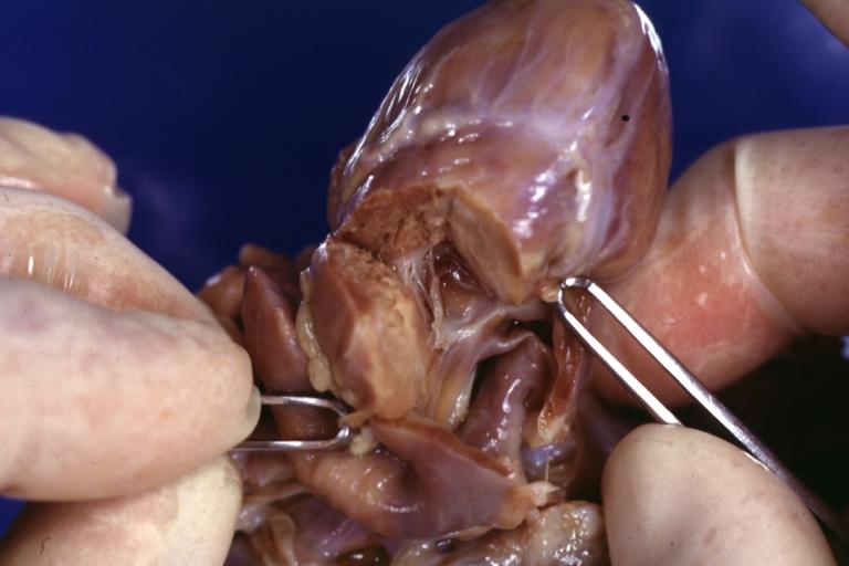
Right Ventricle Hypoplasia: Gross natural color good example showing tiny tricuspid inlet and very small but quite thick right ventricle
-
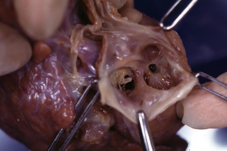
Right Ventricle Hypoplasia: Gross natural color view from right atrium showing patent foramen ovale and very small tricuspid valve
-
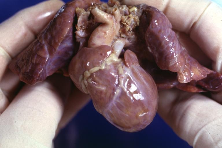
Right Ventricle Hypoplasia: Gross natural color external view of heart showing very large left ventricle and very small right ventricle delineated by anterior descending branch of left coronary artery


External links
- Cleveland Clinic Webchat - Adult Congenital Heart Disease Webchat with Dr. Richard Krasuski.
- Cleveland Clinic Webchat - Adult Congenital Heart Disease Surgery Webchat with Dr. Gosta Pettersson.
- It's My Heart Advocating for and Supporting those affected by Congenital Heart Defects - US Non-Profit under section 501(c)3.
- Saving Little Hearts
- Card-AG, The Cardiologycal Working Group of the University Pediatric Clinic Munster
- The Heart Chest
- American Heart Association
- Congenital Heart Defect
- Treating Congenital Heart Disease
- Coping with Congenital Heart Disease
- Fetal Treatment for Congenital Heart Disease (UCSF Fetal Treatment Center)
- EACTS Congenital Database ― European database of cardiothoracic surgeries with publicly available reports
- Adult Congenital Heart Association
- Congenital Heart Information Network
- Fixing Tiny Tickers - Fetal heart surgery
- Tiny Tickers - Antenatal congenital heart disease information
- Cardiacdiseases.org
Sources
- The ACC/AHA/HRS 2008 Guidelines for Device-Based Therapy of Cardiac Rhythm Abnormalities [1]
References
1. “The Heart Chest.” Non-profit Organization.
- ↑ Epstein AE, DiMarco JP, Ellenbogen KA, Estes NAM III, Freedman RA, Gettes LS, Gillinov AM, Gregoratos G, Hammill SC, Hayes DL, Hlatky MA, Newby LK, Page RL, Schoenfeld MH, Silka MJ, Stevenson LW, Sweeney MO. ACC/AHA/HRS 2008 guidelines for device-based therapy of cardiac rhythm abnormalities: executive summary: a report of the American College of Cardiology/American Heart Association Task Force on Practice Guidelines (Writing Committee to Revise the ACC/AHA/NASPE 2002 Guideline Update for Implantation of Cardiac Pacemakers and Antiarrhythmia Devices). Circulation. 2008; 117: 2820–2840. PMID 18483207
de:Herzfehler lv:Iedzimtās sirds slimības nn:Medfødd hjartefeil sr:Урођене срчане мане uk:Вроджені вади серця wa:Maladeye des bleus påpåds
Overview
Epidemiology and Demographics
Anatomy
Pathophysiology
Genetics
Diagnosis
Clinical Features | Physical Examination | Electrocardiogram | Chest X Ray | Echocardiogram | Cardiac Catheterization
Treatment
Surgical technique for Repair of Perimembranous VSD
Post-operative Treatment
References
External Links for VSD
- Pediatric Heart Surgery
- The Congenital Heart Surgery Video Project
- VSD Repair, Perimembranous Ventricular Septal Defect
- VSD Repair Powerpoint™ Presentation
Acknowledgements and Initial Contributors to Page
Leida Perez, M.D. Redmond Burke M.D.