Thymoma CT: Difference between revisions
Amr Marawan (talk | contribs) No edit summary |
Amr Marawan (talk | contribs) No edit summary |
||
| Line 11: | Line 11: | ||
When a thymic mass is identified, the made is achieved with [[histology]] (obtaining a tissue sample of the mass). When a thymoma is suspected, a [[Computed axial tomography|CT/CAT scan]] is generally performed to estimate the size of the tumor, and can be [[biopsy|biopsied]] with a CT-guided needle. There is a small risk of [[pneumomediastinum]], [[mediastinitis]] and the risk of damaging the [[heart]] or large blood vessels. The final diagnosis is made by removing the thymus. [[Anatomical pathology|Pathological]] investigation of the specimen will reveal if the tumor was benign or malignant, although the initial biopsy is usually indicative.<ref name="pmid10561285">{{cite journal |author=Thomas CR, Wright CD, Loehrer PJ |title=Thymoma: state of the art |journal=[[Journal of Clinical Oncology : Official Journal of the American Society of Clinical Oncology]] |volume=17 |issue=7 |pages=2280–9 |year=1999 |month=July |pmid=10561285 |doi= |url=http://www.jco.org/cgi/pmidlookup?view=long&pmid=10561285 |accessdate=2012-01-18}}</ref> | When a thymic mass is identified, the made is achieved with [[histology]] (obtaining a tissue sample of the mass). When a thymoma is suspected, a [[Computed axial tomography|CT/CAT scan]] is generally performed to estimate the size of the tumor, and can be [[biopsy|biopsied]] with a CT-guided needle. There is a small risk of [[pneumomediastinum]], [[mediastinitis]] and the risk of damaging the [[heart]] or large blood vessels. The final diagnosis is made by removing the thymus. [[Anatomical pathology|Pathological]] investigation of the specimen will reveal if the tumor was benign or malignant, although the initial biopsy is usually indicative.<ref name="pmid10561285">{{cite journal |author=Thomas CR, Wright CD, Loehrer PJ |title=Thymoma: state of the art |journal=[[Journal of Clinical Oncology : Official Journal of the American Society of Clinical Oncology]] |volume=17 |issue=7 |pages=2280–9 |year=1999 |month=July |pmid=10561285 |doi= |url=http://www.jco.org/cgi/pmidlookup?view=long&pmid=10561285 |accessdate=2012-01-18}}</ref> | ||
<gallery> | |||
Image:T001.png| CT shows [[Thymoma]] ( CT scan of the chest revealing a large necrotic mass in the left anterior mediastinum (later proven to be a thymoma) and bilateral hilar lymphadenopathy (from concurrent sarcoidosis) | |||
Image:T002.png| CT shows [[Thymoma]] (Thymoma, Stage IIA. CT scan, axial) | |||
Image:Thymoma-001.jpg| CT shows [[Thymoma]] <small>(Image courtesy of RadsWiki and copylefted) | |||
</gallery> | |||
==References== | ==References== | ||
Revision as of 16:38, 19 February 2014
|
Thymoma Microchapters |
|
Diagnosis |
|---|
|
Case Studies |
|
Thymoma CT On the Web |
|
American Roentgen Ray Society Images of Thymoma CT |
Editor-In-Chief: C. Michael Gibson, M.S., M.D. [1] Associate Editor(s)-in-Chief: Amr Marawan, M.D. [2]
Please help WikiDoc by adding more content here. It's easy! Click here to learn about editing.
Overview
The tumor is generally located inside the thymus, and can be calcified. Increased vascular enhancement can be indicative of malignancy, as can be pleural deposits.
CT Scan
When a thymic mass is identified, the made is achieved with histology (obtaining a tissue sample of the mass). When a thymoma is suspected, a CT/CAT scan is generally performed to estimate the size of the tumor, and can be biopsied with a CT-guided needle. There is a small risk of pneumomediastinum, mediastinitis and the risk of damaging the heart or large blood vessels. The final diagnosis is made by removing the thymus. Pathological investigation of the specimen will reveal if the tumor was benign or malignant, although the initial biopsy is usually indicative.[1]
-
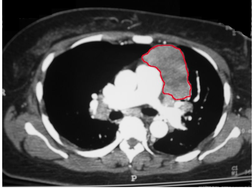
CT shows Thymoma ( CT scan of the chest revealing a large necrotic mass in the left anterior mediastinum (later proven to be a thymoma) and bilateral hilar lymphadenopathy (from concurrent sarcoidosis)
-
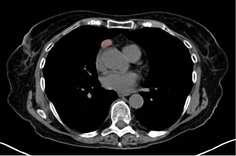
CT shows Thymoma (Thymoma, Stage IIA. CT scan, axial)
-
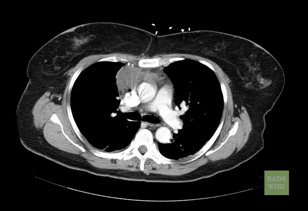
CT shows Thymoma (Image courtesy of RadsWiki and copylefted)


References
- ↑ Thomas CR, Wright CD, Loehrer PJ (1999). "Thymoma: state of the art". Journal of Clinical Oncology : Official Journal of the American Society of Clinical Oncology. 17 (7): 2280–9. PMID 10561285. Retrieved 2012-01-18. Unknown parameter
|month=ignored (help)