TIMI myocardial perfusion grade 0: Difference between revisions
No edit summary |
No edit summary |
||
| (12 intermediate revisions by 3 users not shown) | |||
| Line 1: | Line 1: | ||
{{ | __NOTOC__ | ||
{{Coronary angiography2}} | |||
{{CMG}} | {{CMG}} | ||
| Line 8: | Line 8: | ||
*Intensity Range: 0 | *Intensity Range: 0 | ||
==Example== | |||
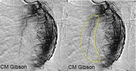
Shown below are a static image and an animated image depicting TIMI myocardial perfusion grade 0. The right hand side of both the static and the moving image shows a crescent shaped dark gray area consistent with normal myocardial perfusion of the posterior wall or the circumflex artery territory. In contrast, note that there is little or no corresponding perfusion in the left hand side of the screen in the territory of the left anterior descending artery. Outlined in yellow is the territory that would ordinarily be perfused by the left anterior descending artery and would be as dark as the perfusion in the circumflex territory. | |||
[[File:TMPG_0_case_01_static.gif|300px]] | |||
[[File:TMPG-0-case-01.gif|300px]] | |||
== | ==Additional Examples== | ||
Click '''[[TIMI myocardial perfusion grade 0 case studies|here]]''' for other examples of TMPG 0. | |||
==References== | |||
{{Coronary Angiography}} | {{Coronary Angiography}} | ||
Latest revision as of 15:26, 13 November 2013
|
Coronary Angiography | |
|
General Principles | |
|---|---|
|
Anatomy & Projection Angles | |
|
Normal Anatomy | |
|
Anatomic Variants | |
|
Projection Angles | |
|
Epicardial Flow & Myocardial Perfusion | |
|
Epicardial Flow | |
|
Myocardial Perfusion | |
|
Lesion Complexity | |
|
ACC/AHA Lesion-Specific Classification of the Primary Target Stenosis | |
|
Lesion Morphology | |
Editor-In-Chief: C. Michael Gibson, M.S., M.D. [1]
Overview
Failure of dye to enter the microvasculature, indicating a lack of tissue level perfusion.
- Intensity Range: 0
Example
Shown below are a static image and an animated image depicting TIMI myocardial perfusion grade 0. The right hand side of both the static and the moving image shows a crescent shaped dark gray area consistent with normal myocardial perfusion of the posterior wall or the circumflex artery territory. In contrast, note that there is little or no corresponding perfusion in the left hand side of the screen in the territory of the left anterior descending artery. Outlined in yellow is the territory that would ordinarily be perfused by the left anterior descending artery and would be as dark as the perfusion in the circumflex territory.
Additional Examples
Click here for other examples of TMPG 0.

