| Lipomatous tumor
|
Age of onset
|
Gender preponderance
|
Location
|
Clinical features
|
Pathologic appearance
|
Other features
|
Pathologic view
|
| Angiolipoma
|
- Second and third decades of life
|
|
- More commonly seen in forearm
- May also affect trunk and upper arm
|
- Subcutaneous nodule
- Tender to palpation
- Less than 2 cm
|
- Encapsulated, yellow nodules with a reddish tinge
- A combination of fatty tissue and vascular channels
- Fibrin thrombi is present in vascular channels (characteristic finding)
|
|
 Contributed by Dr. Dharam Ramnani in Webpathology Contributed by Dr. Dharam Ramnani in Webpathology
|
| Myolipoma
|
- Fifth and sixth decades of life
|
|
- More commonly seen in retroperitoneum, abdomen, pelvis, inguinal region, or abdominal wall
- May also affect extremities
|
- Subcutaneous mass which may also engage superficial muscular fascia
- Size differs depending on the location
|
- Partially encapsulated mass with partially yellow-white cut surface
- A combination of mature adipocytes and sheets of well-differentiated smooth muscle
- No nuclear atypia
- Sieve-like appearance at low magnification (due to interspersed location of smooth muscle component)
|
- Benign
- It is usually large and located in the deep soft tissues
|
 Contributed by Dr. Dharam Ramnani in Webpathology Contributed by Dr. Dharam Ramnani in Webpathology
|
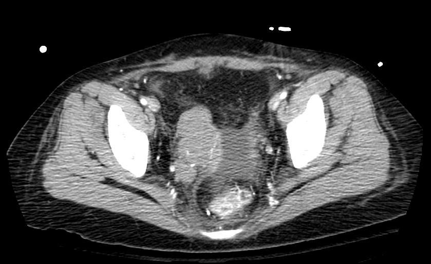
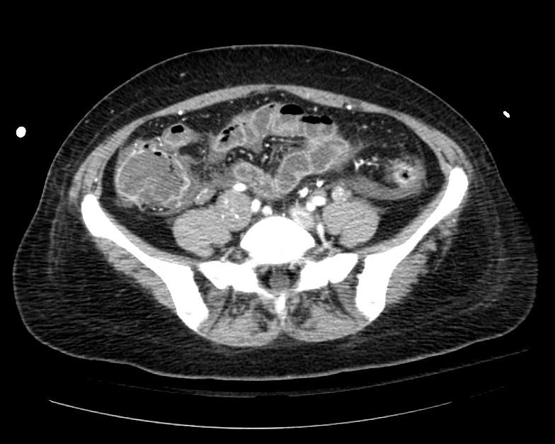
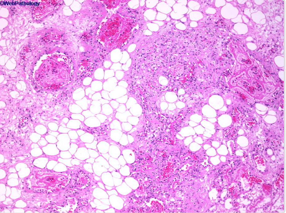
| Myelolipoma
|
|
|
- More commonly seen in adrenal glands
- Other possible locations include:
- thoracic, retroperitoneum and presacral region, mediastinum, liver, and bone
|
- Usually asymptomatic
- May cause abdominal pain, nausea, and constipation (depending on the location and size)
- Uncommonly, may cause retro-peritoneal hemorrhage
- 3 to 7 cm
|
- A combination of bone marrow elements and adipose tissue in varying proportions
- May show myxoid changes
|
- Well-circumscribed radiolucent mass in radiologic imaging
- May have hromonal activity
|
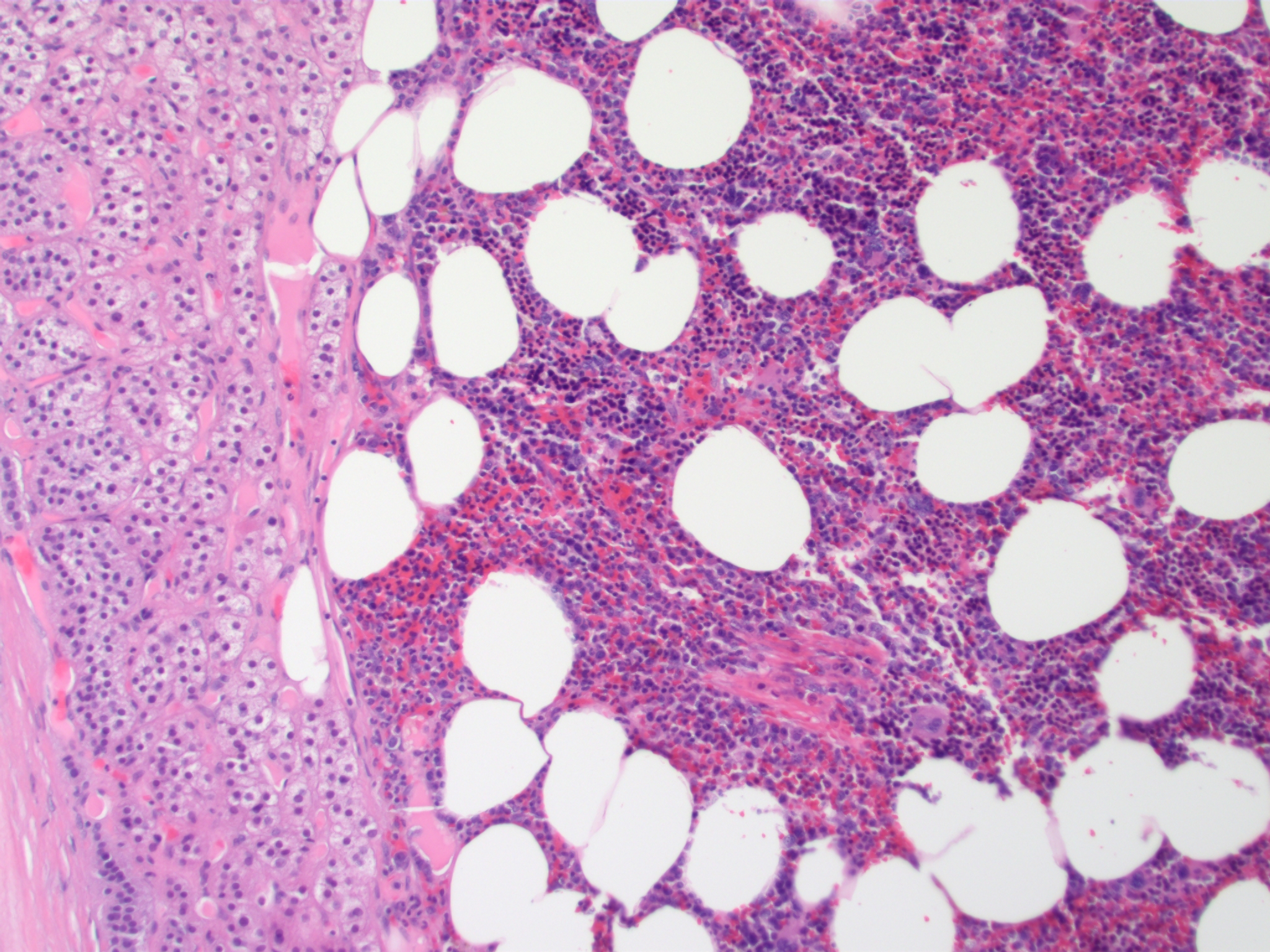 Contributed by Sarahkayb in Wikimedia commons Contributed by Sarahkayb in Wikimedia commons
|
| Spindle Cell/Pleomorphic Lipoma
|
- Fifth to seventh decades of life
|
|
- More commonly seen in posterior neck, shoulder, and back
- It is also reported in oral cavity
|
- Subcutaneous nodule with firm consistency
- Slowly growing and painless
- Mostly between 3 to 5 cm
|
- Similar to ordinary lipoma
- A combination of mature fat cells and spindle cell or pleomorphic elements
- Lipomatous component may vary in amount
|
- Immunohistochemically positive for CD34
- Benign
|
 Contributed by Nephron in Wikimedia commons Contributed by Nephron in Wikimedia commons
|
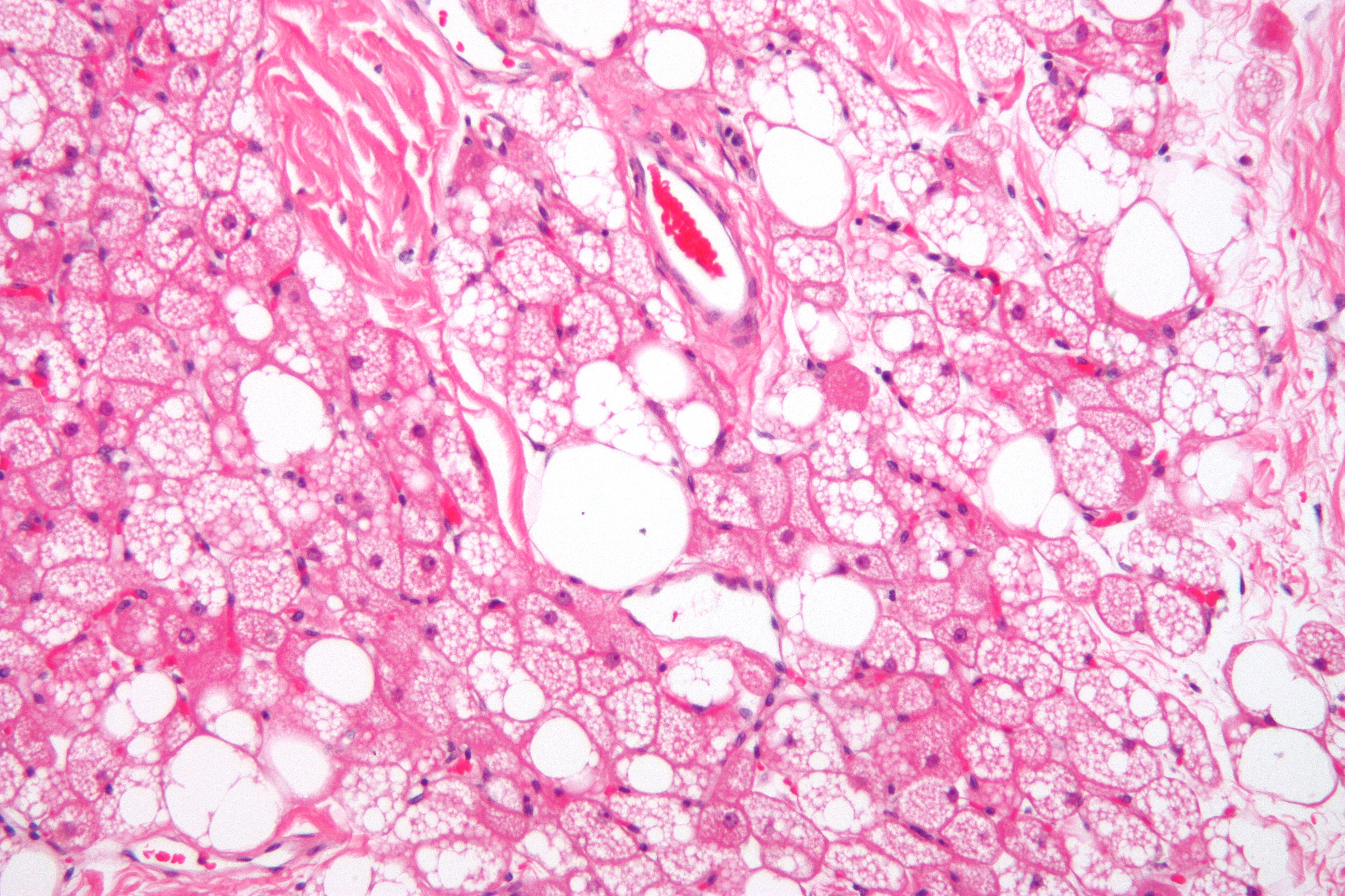
| Chondroid Lipoma
|
- Third or fourth decade of life
|
|
- More commonly seen in limbs and limb girdles
- May also involve trunk, and the head and neck region, particularly the oral cavity
|
- Slowly growing painless mass
- Sizes ranges from 1 to 11 cm
|
- Encapsulated tumor with a yellow, white, or pink-tan cut surface
- A combination of mature adipocytes in association with nests of vacuolated cells in a myxochondroid or hyalinized fibrous background
|
- Heterogeneous soft tissue mass in radiologic imaging
- Benign
|
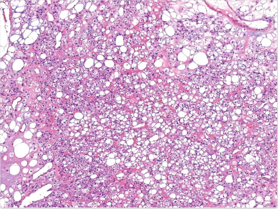 Contributed by Dr. Dharam Ramnani in Webpathology Contributed by Dr. Dharam Ramnani in Webpathology
|
| Hibernoma
|
|
|
- Most commonly seen in thigh
- May also affect shoulder, back, neck, chest, arm, and abdominal cavity/retroperitoneum
|
- Slowly growing, painless, subcutaneous mass
- Affects intramuscular in 10% of the cases
- Size varies between 5 to 15 cm
|
- Well-defined, soft, and mobile mass
- A combination of vacuolated granular eosinophilic cells with abundant mithochondria and high vascular content
|
- Immunohistochemically positive for S-100
- Benign
|
 Contributed by Nephron in Wikimedia commons Contributed by Nephron in Wikimedia commons
|
| Intramuscular and Intermuscular Lipomas
|
- Fourth to seventh decades of life
|
|
- Most commonly seen in large muscles of the extremities, especially in thigh, shoulder, and upper arm
|
- Painless, slowly growing mass
- Visible during muscle contraction
|
- Infiltrative adipose thissue within the muscle
- Absence of nuclear atypia
|
- May be very small or more than 20 cm
|
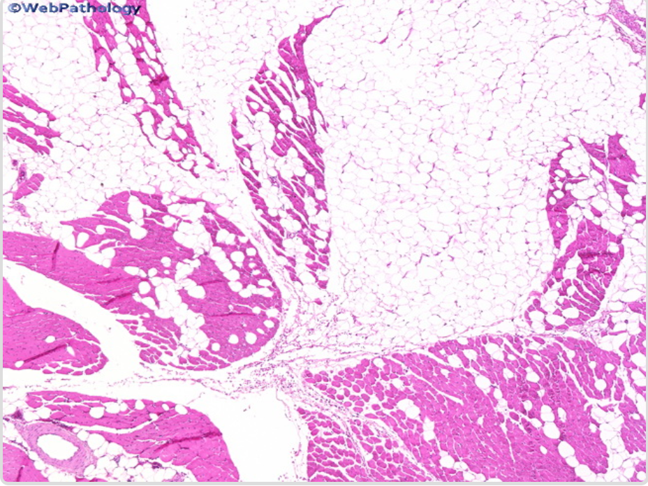 Contributed by Dr. Dharam Ramnani in Webpathology Contributed by Dr. Dharam Ramnani in Webpathology
|
| Lipomas of Tendon Sheaths and Joints
|
- Second and third decades of life
|
- Female = male
- Lipoma of joints affects men more frequently than women
|
- Most commonly seen in wrist and hand
- May also affect ankle and foot
- Bilateral and symmetric location in 50% of the cases
|
- May cause severe pain, trigger finger, or even symptoms of carpal tunnel syndrome
|
- May have 2 different shape:
- A single adipose tissue extending along the tendon sheet
- A lipomatous lesion composed mostly from hypertrophic synovial villi
|
- Radiologic imaging may show a lesion of less density than the surrounding tissue
|
_
|
| Lumbosacral Lipoma
|
|
|
- It occurs in the lumbosacral region and overlies the spine
|
- Initially, asymptomatic
- Later signs and symptoms of progressive myelopathy or radiculopathy in the lower legs, bladder, or bowel
|
- Uncapsulated mass that consists of lobulated adipose tissue
- Vascular proliferation and smooth muscle tissue may be present
|
- Almost always associated with spina bifida or a similar laminar defect (lipomyeloschisis)
|
_
|
| Neural Fibrolipoma (Lipofibromatous Hamartoma of Nerves)
|
- First three decades of life
|
- Female > male (in the presence of macrodactyly)
|
- Most commonly affect median nerve and its branches
- May also affect ulnar, radial, peroneal, and cranial nerves
|
- May cause neuropathy, pain, paresthesia, and decreased sensation
- Carpal tunnel syndrome may also occur
|
- A sausage-shaped mass with soft teture that has diffusely infiltrated a large nerve and its branches
- Fibrofatty tissue surronding and infiltrating the nerve trunk
|
- Overgrowth of bone and macrodactyly of the digits innervated by the affected nerve is seen in one third of the cases
|
_
|