Pyelonephritis echocardiography and ultrasound
|
Pyelonephritis Microchapters |
|
Diagnosis |
|
Treatment |
|
Case Studies |
|
Pyelonephritis echocardiography and ultrasound On the Web |
|
American Roentgen Ray Society Images of Pyelonephritis echocardiography and ultrasound |
|
Risk calculators and risk factors for Pyelonephritis echocardiography and ultrasound |
Editor-In-Chief: C. Michael Gibson, M.S., M.D. [1]; Associate Editor(s)-in-Chief: Usama Talib, BSc, MD [2]
Overview
Ultrasonography is an effective non-invasive technique in the diagnosis of pyelonephritis. It is sometimes used as a replacement of cortical scintigraphy in the diagnosis of acute pyelonephritis in children.[1][2][3]
Ultrasound
The diagnosis of pyelonephritis using ultrasound may feature the following:[4]
- In case of recurrent urinary tract infections, it can help to rule out presence of anatomical defects like polycystic kidney disease.
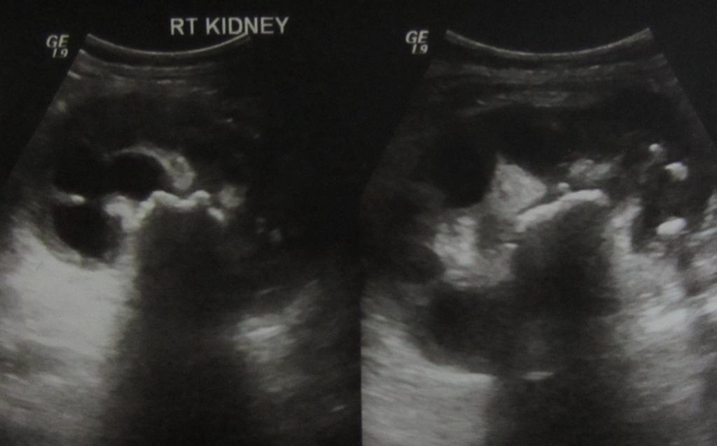
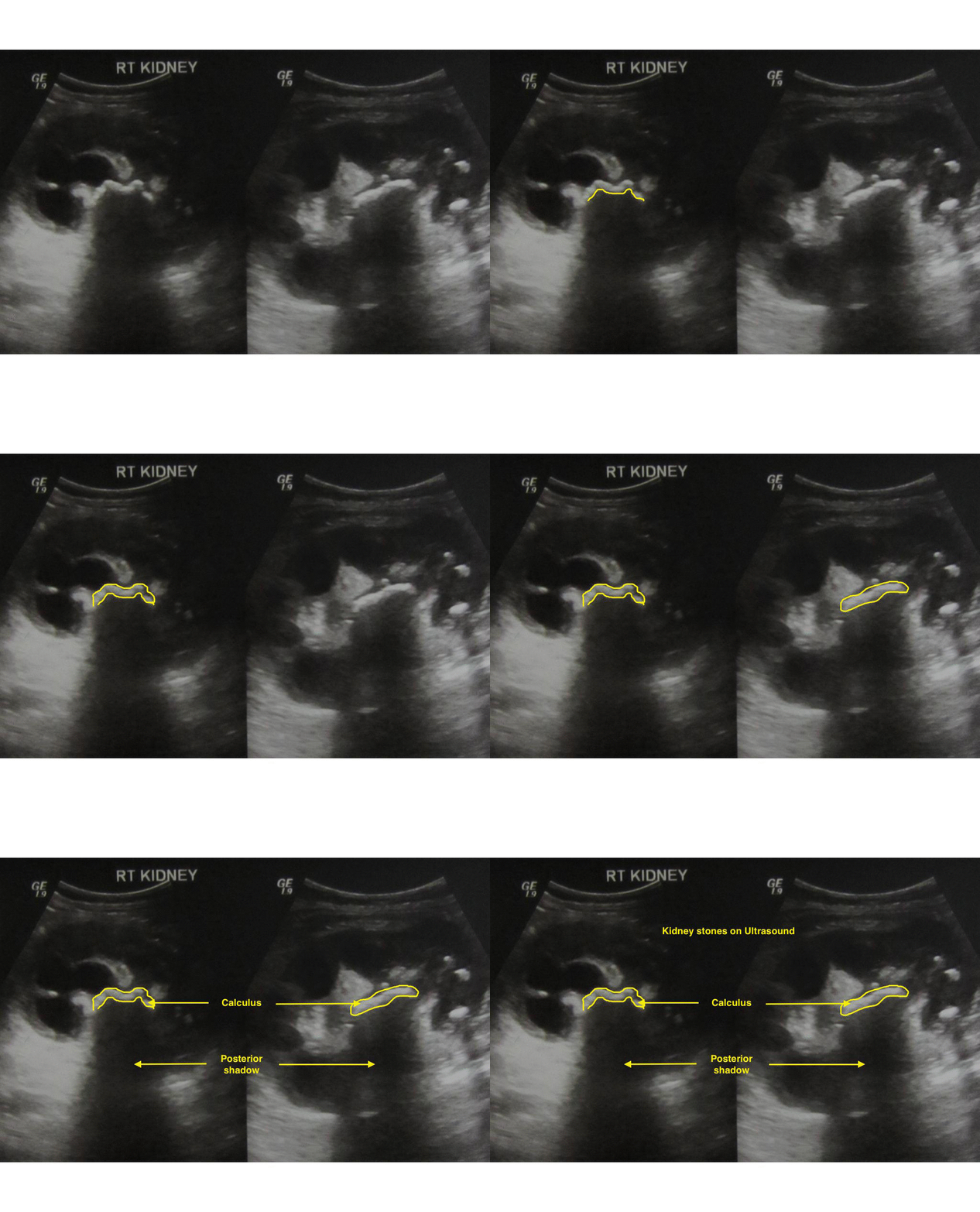
- Emphysematous pyelonephritis: Ultrasound will present an enlarged kidney containing air filled regions within the renal parenchyma, with shadowing shown posterior to the defect; It might not be as helpful in detecting the depth of involvement and stones can also show posterior shadowing.
- Xanthogranulomatous pyelonephritis: Ultrasound shows the inflammatory mass, with central shadowing depicting renal calculi.
References
- ↑ Fowler JE, Perkins T (1994). "Presentation, diagnosis and treatment of renal abscesses: 1972-1988". J Urol. 151 (4): 847–51. PMID 8126807.
- ↑ Gupta K, Hooton TM, Naber KG, Wullt B, Colgan R, Miller LG; et al. (2011). "International clinical practice guidelines for the treatment of acute uncomplicated cystitis and pyelonephritis in women: A 2010 update by the Infectious Diseases Society of America and the European Society for Microbiology and Infectious Diseases". Clin Infect Dis. 52 (5): e103–20. doi:10.1093/cid/ciq257. PMID 21292654.
- ↑ Kawashima A, LeRoy AJ (2003). "Radiologic evaluation of patients with renal infections". Infect Dis Clin North Am. 17 (2): 433–56. PMID 12848478.
- ↑ Radiopaedia.org. Case courtesy of Dr Ian Bickle, <a href="https://radiopaedia.org/">Radiopaedia.org</a>. From the case <a href="https://radiopaedia.org/cases/20872">rID: 20872

