Peutz-Jeghers syndrome pathophysiology: Difference between revisions
Hamid Qazi (talk | contribs) |
|||
| (19 intermediate revisions by 2 users not shown) | |||
| Line 1: | Line 1: | ||
__NOTOC__ | __NOTOC__ | ||
{{Peutz-Jeghers syndrome}} | {{Peutz-Jeghers syndrome}} | ||
{{CMG}} {{AE}} {{HQ}} | {{CMG}}; {{AE}} {{HQ}} | ||
==Overview== | ==Overview== | ||
Peutz-Jeghers syndrome is transmitted in an [[autosomal dominant]] pattern. Polyps of Peutz-Jeghers syndrome are usually non-neoplastic [[hamartomas]]. | Peutz-Jeghers syndrome is transmitted in an [[autosomal dominant]] pattern. [[Polyps]] of Peutz-Jeghers syndrome are usually non-neoplastic [[hamartomas]]. It is thought that Peutz-Jeghers syndrome is the result of [[Deletion (genetics)|deletion]] or [[Deletion (genetics)|partial deletion]] of [[STK11]] (LBK1) gene, located on [[chromosome]] 19p13.3. Mucutaneous [[pigmentation]] ([[macules]]) are caused by [[pigment]]-laden [[macrophages]] in the [[dermis]]. | ||
==Pathophysiology== | ==Pathophysiology== | ||
===Pathogenesis=== | |||
*It is thought that Peutz-Jeghers syndrome is the result of [[Deletion (genetics)|deletion]] or [[Deletion (genetics)|partial deletion]] of [[STK11]] (LBK1) gene, located on [[chromosome]] 19p13.3.<ref name="KopacovaTacheci2009">{{cite journal|last1=Kopacova|first1=Marcela|last2=Tacheci|first2=Ilja|last3=Rejchrt|first3=Stanislav|last4=Bures|first4=Jan|title=Peutz-Jeghers syndrome: Diagnostic and therapeuticapproach|journal=World Journal of Gastroenterology|volume=15|issue=43|year=2009|pages=5397|issn=1007-9327|doi=10.3748/wjg.15.5397}}</ref><ref name="BuckHarned1992">{{cite journal|last1=Buck|first1=J L|last2=Harned|first2=R K|last3=Lichtenstein|first3=J E|last4=Sobin|first4=L H|title=Peutz-Jeghers syndrome.|journal=RadioGraphics|volume=12|issue=2|year=1992|pages=365–378|issn=0271-5333|doi=10.1148/radiographics.12.2.1561426}}</ref> | |||
*[[STK11]] [[protein]] plays an important role in [[second messenger]] [[signal transduction]] and is found to regulate [[cellular]] [[proliferation]], controls [[cell]] polarity, and responds to low energy states. | |||
*In Mammalian studies, [[STK11]] is shown in the inhibition of [[AMP-activated protein kinase]] (AMPK), and signals downstream to inhibit the [[mammalian target of rapamycin]] ([[mTOR]]). | |||
** The [[Mammalian target of rapamycin|mTOR pathway]] is dysregulated in Peutz-Jeghers syndrome. | |||
* Pathogenesis of mucutaneous [[pigmentation]] ([[Macule|macules]]) | |||
**Caused by [[pigment]]-laden [[Macrophage|macrophages]] in the [[dermis]]. | |||
===Genetics=== | ===Genetics=== | ||
*Peutz-Jeghers syndrome is inherited in an [[autosomal dominant]] pattern. | *Peutz-Jeghers syndrome is [[inherited]] in an [[autosomal dominant]] pattern. | ||
=== | |||
* | ==Associated Conditions== | ||
* | Conditions associated with Peutz-Jeghers syndrome include: | ||
*[[Breast Cancer]] | |||
*[[Colorectal cancer|Colon Cancer]] | |||
*[[Pancreatic cancer]] | |||
*[[Ovarian cancer]] | |||
*[[Cervical cancer]] | |||
*[[Testicular cancer]] | |||
==Gross Pathology== | |||
*On gorss pathology, Peutz-Jeghers syndrome associated [[Polyp|polyps]] have a unique [[smooth muscle]] core that arborizes throughout the [[polyp]].<ref name="KopacovaTacheci2009">{{cite journal|last1=Kopacova|first1=Marcela|last2=Tacheci|first2=Ilja|last3=Rejchrt|first3=Stanislav|last4=Bures|first4=Jan|title=Peutz-Jeghers syndrome: Diagnostic and therapeuticapproach|journal=World Journal of Gastroenterology|volume=15|issue=43|year=2009|pages=5397|issn=1007-9327|doi=10.3748/wjg.15.5397}}</ref> | |||
**These [[Polyp|polyps]] can only be differentiated from other [[polyp]] types by [[histopathology]]. | |||
* | ==Microscopic Pathology== | ||
* [[Polyp|Polyps]] of Peutz-Jeghers syndrome are usually non-neoplastic [[hamartomas]].<ref>Pathology of Peutz-Jeghers syndrome. Dr Amir Rezaee and Dr Alexandra Stanislavsky et al. Radiopaedia.org 2015. http://radiopaedia.org/articles/peutz-jeghers-syndrome-2</ref> | |||
* On microscopic histopathological analysis, [[Polyp|polyps]] have the following characteristic findings:<ref name="BuckHarned1992">{{cite journal|last1=Buck|first1=J L|last2=Harned|first2=R K|last3=Lichtenstein|first3=J E|last4=Sobin|first4=L H|title=Peutz-Jeghers syndrome.|journal=RadioGraphics|volume=12|issue=2|year=1992|pages=365–378|issn=0271-5333|doi=10.1148/radiographics.12.2.1561426}}</ref> | |||
* | *Frond-like [[polyp]] with all three components of [[mucosa]]: | ||
** | **Muscosal [[epithelium]] (melanotic [[mucosa]], [[goblet cells]]) | ||
**[[Lamina propria]] | |||
**[[Muscularis mucosae]] | |||
[[File:Peutz-Jeghers syndrome polyp .jpg|none|thumb|260x260px|Peutz-Jeghers Polyp Histology [https://upload.wikimedia.org/wikipedia/commons/c/c6/Peutz-Jeghers_syndrome_polyp.jpg Source: By Nephron (Own work), via Wikimedia Commons]]] | |||
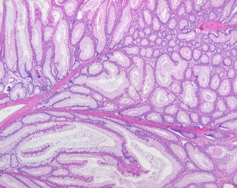
[[File:Colon histology with Peutz-Jeghers polyp.jpg|200x200px|thumb|Peutz-Jeghers Polyp Histology [https://www.wikidoc.org/images/0/03/Colon_histology_with_Peutz-Jeghers_polyp.jpg Source:Libre Pathology]]] | |||
== References == | == References == | ||
{{reflist|2}} | {{reflist|2}} | ||
{{WH}} | |||
{{WS}} | |||
[[Category:Gastroenterology]] | |||
[[Category:Surgery]] | |||
[[Category:Oncology]] | |||
[[Category:Disease]] | |||
[[Category:Pediatrics]] | |||
[[Category:Up-To-Date]] | |||
Latest revision as of 15:16, 21 December 2017
|
Peutz-Jeghers syndrome Microchapters |
|
Diagnosis |
|---|
|
Treatment |
|
Case Studies |
|
Peutz-Jeghers syndrome pathophysiology On the Web |
|
American Roentgen Ray Society Images of Peutz-Jeghers syndrome pathophysiology |
|
Risk calculators and risk factors for Peutz-Jeghers syndrome pathophysiology |
Editor-In-Chief: C. Michael Gibson, M.S., M.D. [1]; Associate Editor(s)-in-Chief: Hamid Qazi, MD, BSc [2]
Overview
Peutz-Jeghers syndrome is transmitted in an autosomal dominant pattern. Polyps of Peutz-Jeghers syndrome are usually non-neoplastic hamartomas. It is thought that Peutz-Jeghers syndrome is the result of deletion or partial deletion of STK11 (LBK1) gene, located on chromosome 19p13.3. Mucutaneous pigmentation (macules) are caused by pigment-laden macrophages in the dermis.
Pathophysiology
Pathogenesis
- It is thought that Peutz-Jeghers syndrome is the result of deletion or partial deletion of STK11 (LBK1) gene, located on chromosome 19p13.3.[1][2]
- STK11 protein plays an important role in second messenger signal transduction and is found to regulate cellular proliferation, controls cell polarity, and responds to low energy states.
- In Mammalian studies, STK11 is shown in the inhibition of AMP-activated protein kinase (AMPK), and signals downstream to inhibit the mammalian target of rapamycin (mTOR).
- The mTOR pathway is dysregulated in Peutz-Jeghers syndrome.
- Pathogenesis of mucutaneous pigmentation (macules)
- Caused by pigment-laden macrophages in the dermis.
Genetics
- Peutz-Jeghers syndrome is inherited in an autosomal dominant pattern.
Associated Conditions
Conditions associated with Peutz-Jeghers syndrome include:
Gross Pathology
- On gorss pathology, Peutz-Jeghers syndrome associated polyps have a unique smooth muscle core that arborizes throughout the polyp.[1]
- These polyps can only be differentiated from other polyp types by histopathology.
Microscopic Pathology
- Polyps of Peutz-Jeghers syndrome are usually non-neoplastic hamartomas.[3]
- On microscopic histopathological analysis, polyps have the following characteristic findings:[2]
- Frond-like polyp with all three components of mucosa:
- Muscosal epithelium (melanotic mucosa, goblet cells)
- Lamina propria
- Muscularis mucosae


References
- ↑ 1.0 1.1 Kopacova, Marcela; Tacheci, Ilja; Rejchrt, Stanislav; Bures, Jan (2009). "Peutz-Jeghers syndrome: Diagnostic and therapeuticapproach". World Journal of Gastroenterology. 15 (43): 5397. doi:10.3748/wjg.15.5397. ISSN 1007-9327.
- ↑ 2.0 2.1 Buck, J L; Harned, R K; Lichtenstein, J E; Sobin, L H (1992). "Peutz-Jeghers syndrome". RadioGraphics. 12 (2): 365–378. doi:10.1148/radiographics.12.2.1561426. ISSN 0271-5333.
- ↑ Pathology of Peutz-Jeghers syndrome. Dr Amir Rezaee and Dr Alexandra Stanislavsky et al. Radiopaedia.org 2015. http://radiopaedia.org/articles/peutz-jeghers-syndrome-2