|
|
| (41 intermediate revisions by 7 users not shown) |
| Line 1: |
Line 1: |
| {{Infobox_Disease
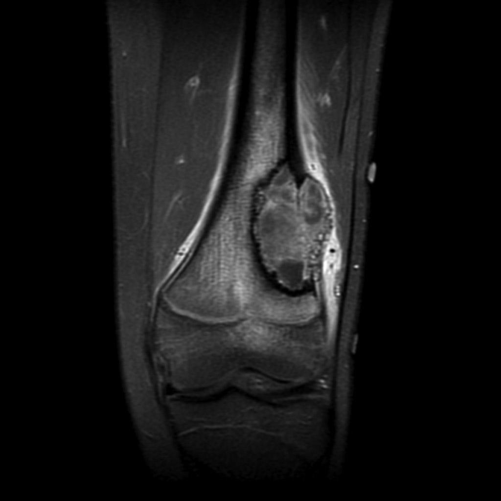
| | [[File:Osteosarcoma-distal-femur (2).jpg|thumb|Conventional Osteosarcoma of the distal femur of an 18 year old male.Case courtesy of A.Prof Frank Gaillard, <a href="https://radiopaedia.org/">Radiopaedia.org</a>. From the case <a href="https://radiopaedia.org/cases/7621">rID: 7621</a>]] |
| | Name = Osteosarcoma
| | __NOTOC__ |
| | DiseasesDB = 9392
| |
| | ICD10 = {{ICD10|C|40||c|40}}-{{ICD10|C|41||c|40}}
| |
| | ICD9 = {{ICD9|170}}
| |
| | ICDO = {{ICDO|9180|3}}
| |
| | OMIM = 259500
| |
| | MedlinePlus = 001650
| |
| | MeshID = D012516
| |
| }}
| |
| {{Osteosarcoma}} | | {{Osteosarcoma}} |
| '''For patient information click [[{{PAGENAME}} (patient information)|here]]''' | | '''For patient information click [[{{PAGENAME}} (patient information)|here]]''' |
|
| |
|
| {{CMG}} | | {{CMG}}; {{AE}}[[User:DrMars|Mohammadmain Rezazadehsaatlou[2]]]. |
| | |
| '''Associate Editor-In-Chief:''' {{CZ}}
| |
| | |
| {{Editor Help}}
| |
|
| |
|
| | {{SK}} Osteogenic sarcoma; Osteosarcoma; Osteoblastic osteosarcoma; Osteoblastic sarcoma; Osteochondrosarcoma; Telangiectatic osteosarcoma; Small-cell osteosarcoma; Small cell osteosarcoma; Conventional central osteosarcoma; Conventional osteosarcoma; Intraosseous low-grade osteosarcoma; Low grade osteosarcoma; Low-grade osteosarcoma; Intraosseous osteosarcoma; Parosteal osteosarcoma; Periosteal osteosarcoma; Extraskeletal osteosarcoma; High-grade osteosarcoma; High grade osteosarcoma |
| ==[[Osteosarcoma overview|Overview]]== | | ==[[Osteosarcoma overview|Overview]]== |
|
| |
|
| ==[[Osteosarcoma historical perspective|Historical Perspective]]== | | ==[[Osteosarcoma historical perspective|Historical Perspective]]== |
| | |
| | ==[[Osteosarcoma classification|Classification]]== |
|
| |
|
| ==[[Osteosarcoma pathophysiology|Pathophysiology]]== | | ==[[Osteosarcoma pathophysiology|Pathophysiology]]== |
|
| |
|
| ==[[Osteosarcoma epidemiology and demographics|Epidemiology & Demographics]]== | | ==[[Osteosarcoma causes|Causes]]== |
| | |
| | ==[[Osteosarcoma differential diagnosis|Differentiating Osteosarcoma from other Diseases]]== |
| | |
| | ==[[Osteosarcoma epidemiology and demographics|Epidemiology and Demographics]]== |
|
| |
|
| ==[[Osteosarcoma risk factors|Risk Factors]]== | | ==[[Osteosarcoma risk factors|Risk Factors]]== |
| Line 30: |
Line 25: |
| ==[[Osteosarcoma screening|Screening]]== | | ==[[Osteosarcoma screening|Screening]]== |
|
| |
|
| ==[[Osteosarcoma causes|Causes]]==
| | ==[[Osteosarcoma natural history|Natural History, Complications and Prognosis]]== |
| | |
| ==[[Osteosarcoma differential diagnosis|Differentiating Osteosarcoma]]==
| |
| | |
| ==[[Osteosarcoma natural history|Complications & Prognosis]]== | |
|
| |
|
| ==Diagnosis== | | ==Diagnosis== |
| [[Osteosarcoma history and symptoms|History and Symptoms]] | [[Osteosarcoma physical examination|Physical Examination]] | [[Osteosarcoma staging|Staging]] | [[Osteosarcoma laboratory tests|Laboratory tests]] | [[Osteosarcoma electrocardiogram|Electrocardiogram]] | [[Osteosarcoma x ray|X Rays]] | [[Osteosarcoma CT|CT]] | [[Osteosarcoma MRI|MRI]] [[Osteosarcoma echocardiography or ultrasound|Echocardiography or Ultrasound]] | [[Osteosarcoma other imaging findings|Other images]] | [[Osteosarcoma other diagnostic studies|Alternative diagnostics]] | | [[Osteosarcoma staging|Staging]] | [[Osteosarcoma history and symptoms|History and Symptoms]] | [[Osteosarcoma physical examination|Physical Examination]] | [[Osteosarcoma laboratory findings|Laboratory Findings]] | [[Osteosarcoma biopsy|Biopsy]] | [[Osteosarcoma x ray|X Ray]] | [[Osteosarcoma CT|CT]] | [[Osteosarcoma MRI|MRI]] | [[Osteosarcoma other imaging findings|Other Imaging Findings]] | [[Osteosarcoma other diagnostic studies|Other Diagnostic Studies]] |
|
| |
|
| ==Treatment== | | ==Treatment== |
| [[Osteosarcoma medical therapy|Medical therapy]] | [[Osteosarcoma surgery|Surgical options]] | [[Osteosarcoma primary prevention|Primary prevention]] | [[Osteosarcoma secondary prevention|Secondary prevention]] | [[Osteosarcoma cost-effectiveness of therapy|Financial costs]] | [[Osteosarcoma future or investigational therapies|Future therapies]] | | [[Osteosarcoma medical therapy|Medical Therapy]] | [[Osteosarcoma surgery|Surgery]] | [[Osteosarcoma prevention|Prevention]] | [[Osteosarcoma cost-effectiveness of therapy|Cost-Effectiveness of Therapy]] | [[Osteosarcoma future or investigational therapies|Future or Investigational Therapies]] |
| | |
| ==Symptoms==
| |
| Many patients first complain of pain that may be worse at night, and may have been occurring for some time. If the tumour is large, it can appear as a swelling. The affected bone is not as strong as normal bones and may fracture with minor trauma (a pathological fracture).
| |
| | |
| *Bone fracture (may occur after what seems like a routine movement)
| |
| *Bone pain
| |
| *Limitation of motion
| |
| *Limping (if the tumor is in the leg)
| |
| *Pain when lifting (if the tumor is in the arm)
| |
| *Tenderness, swelling, or redness at the site of the tumor
| |
| | |
| ==Genetics==
| |
| | |
| Hereditary syndromes of osteosarcoma have been identified<ref>Wang LL. Biology of osteogenic sarcoma. Cancer J 11:294-305, 2005.</ref>:
| |
| *[[Rothmund-Thomson Syndrome]]
| |
| *RECQL4 gene mutations
| |
| *[[RB1]] gene mutations (also implicated in [[retinoblastoma]])
| |
| *[[Li-Fraumeni syndrome]]
| |
| These syndromes are extremely rare within the Osteosarcoma diagnosis, and probably represent less than 0.5% of those diagnosed
| |
| | |
| ==Diagnosis==
| |
| | |
| Family [[physicians]] and orthopedists rarely see a [[malignant]] bone tumor (most bone tumors are [[benign]]). Thus, many patients are initially misdiagnosed with [[cysts]] or muscle problems, and some are sent straight to [[physical therapy]] without an [[x-ray]].
| |
| | |
| The route to [[osteosarcoma]] diagnosis usually begins with an [[x-ray]], continues with a combination of scans ([[CT scan]], [[PET scan]], bone scan, [[MRI]]) and ends with a surgical [[biopsy]]. Much can be seen on films, but the [[biopsy]] is the only definitive proof that a bone tumor is indeed [[malignant]] or [[benign]].
| |
| | |
| The biopsy of suspected [[osteosarcoma]] should be performed by a qualified orthopedic [[oncologist]]. The [[American Cancer Society]] states: "Probably in no other [[cancer]] is it as important to perform this procedure properly. An improperly performed [[biopsy]] may make it difficult to save the affected limb from [[amputation]]."
| |
| | |
| ===MRI===
| |
| | |
| [http://www.radswiki.net Images courtesy of RadsWiki]
| |
| | |
| <gallery perRow="3">
| |
| Image:Osteosarcoma-001.jpg|Plain film: Osteosarcoma
| |
| Image:Osteosarcoma-002.jpg|Plain film: Osteosarcoma
| |
| Image:Osteosarcoma-003.jpg|Bone scan
| |
| Image:Osteosarcoma-004.jpg|MRI T1
| |
| Image:Osteosarcoma-005.jpg|MRI T1
| |
| Image:Osteosarcoma-006.jpg|MRI T1 post gad
| |
| Image:Osteosarcoma-007.jpg|MRI T1 post gad
| |
| Image:Osteosarcoma-008.jpg|MRI T2 fat sat
| |
| Image:Osteosarcoma-009.jpg|MRI T2 fat sat
| |
| </gallery>
| |
| | |
| ==Treatment==
| |
| | |
| Patients with osteosarcoma are best managed by a medical oncologist and an orthopedic oncologist experienced in managing [[sarcomas]]. Current standard treatment is to use [[neoadjuvant]] [[chemotherapy]] ([[chemotherapy]] given before [[surgery]]) followed by surgical resection. The percentage of tumor cell [[necrosis]] (cell death) seen in the tumor after surgery gives an idea of the prognosis and also lets the oncologist know if the [[chemotherapy]] regime should be altered after surgery.
| |
| | |
| Standard therapy is a combination of limb-salvage [[orthopedic surgery]] and a combination of high dose [[methotrexate]] with [[leucovorin]] rescue, intra-arterial [[cisplatin]] [[caffeine]], [[adriamycin]], [[ifosfamide]] with [[mesna]], BCD, [[etoposide]], muramyl tri-peptite (MTP).
| |
| | |
| Ifosfamide can be used as an adjuvant treatment if the [[necrosis]] rate is low.
| |
| | |
| 3-year event free survival ranges from 50% to 75%. and 5-year survival ranges from
| |
| 60% to 85+% in some studies. Overall, 60-65% treated 5-years ago (2000) will be alive today.
| |
| Osteosarcoma has one of the lowest survival rates for pediatric cancer despite chemotherapy's success in osteosarcoma of 6 chemotherapies, [[interferon-alpha]], [[interleukin-2]], and being the prototype
| |
| of solid tumors in cancer.
| |
| | |
| Treatment studies come from Children's hospital Boston, Memorial Sloan-Kettering, Children's Oncology Group, Italian Oncology Group, Japan, and MD Anderson in Texas.
| |
| | |
| Fluids are given for hydration.
| |
| | |
| Drugs like [[Kytril]] and [[Zofran]] help with [[nausea]] and [[vomiting]].
| |
| | |
| [[Neupogen]], [[epogen]], [[Neulasta]] help with [[white blood cell]] counts and [[neutrophil]] counts.
| |
| | |
| Blood helps with [[anemia]].
| |
| | |
| ==Prognosis==
| |
| | |
| *Prognosis is separated into three groups.
| |
| | |
| * '''Stage I''' osteosarcoma is rare and includes parosteal osteosarcoma or low-grade central osteosarcoma. It has an excellent prognosis (>90%) with wide resection.
| |
| | |
| * '''Stage IIb''' prognosis depends on the site of the tumor (proximal tibia, femur, pelvis, etc.) size of the tumor mass (in cm.), the degree of necrosis from neoadjuvant chemotherapy (beforeoperation chemotherapy), and pathological factors like the degree of p-glycoprotein, whether your tumor is cxcr4-positive.<ref>http://www.osteosarcomasupport.org/cxcr4_metastases.pdf</ref> Her2-positive as these can lead to distant metastases to the lung. Longer time to metastases, more than 12 months or 24 months and the number of metastases and resectability of them lead to the best prognosis with metastatic osteosarcoma. It is better to have fewer metastases than longer time to metastases. Those with a longer length of time(>24months) and few nodules (2 or fewer) have the best prognosis with a 2-year survival after the metastases of 50% 5-year of 40% and 10 year 20%. If metastases are both local and regional the prognosis is different unfortunately.
| |
| | |
| * Initial Presentation of '''stage III''' osteosarcoma with lung metastates depends on the resectability of the primary tumor and lung nodules, degree of necrosis of the primary tumor, and maybe the number of metastases. Overall prognosis is 30% or greater depending.
| |
| | |
| ==Case Examples==
| |
| | |
| ===Case #1===
| |
| | |
| ====Clinical Summary====
| |
| | |
| This 14-year-old white male first experienced mild pain in the left knee after playing baseball, approximately two months prior to admission. The pain persisted in an intermittent fashion, and was described as being somewhat worse at night. Approximately two weeks prior to admission, the pain increased significantly and was accompanied by marked swelling and loss of considerable motion of the knee joint. These symptoms were accompanied by a history of decreased appetite, lethargy, and a 10-pound weight loss. On physical examination, the left knee was enlarged diffusely, firm, and non-tender. Following biopsy, the patient was subjected to surgical removal of the distal femur and knee with placement of a prosthetic knee joint and bone grafts.
| |
| | |
| ====Autopsy Findings====
| |
| | |
| The distal diaphysis of the femur and adjacent soft tissues were involved in a 15 x 10 x 10-cm mass. The cut surface of the mass was fleshy white, with focal areas of hemorrhage.
| |
| | |
| ====Pathological Findings====
| |
| | |
| [http://www.peir.net Image courtesy of Professor Peter Anderson DVM PhD and published with permission © PEIR, University of Alabama at Birmingham, Department of Pathology]
| |
| | |
| [[Image:Osteosarcoma case 001.jpg|left|thumb|300px|This is a photograph of the patient prior to surgery. Note the marked swelling of the knee.]]
| |
| <br clear="left"/>
| |
| | |
| [[Image:Osteosarcoma case 002.jpg|left|thumb|300px|This is a radiograph showing the tumor in the distal femur.]]
| |
| <br clear="left"/>
| |
| | |
| [[Image:Osteosarcoma case 003.jpg|left|thumb|300px|This is another view of the tumor in the distal femur.]]
| |
| <br clear="left"/>
| |
|
| |
|
| [[Image:Osteosarcoma case 004.jpg|left|thumb|300px|This is a gross photograph of the surgical specimen with tissue dissected away to demonstrate the tumor mass.]]
| | ==Case Studies== |
| <br clear="left"/>
| | :[[Osteosarcoma case study one|Case #1]] |
| | |
| [[Image:Osteosarcoma case 005.jpg|left|thumb|300px|These are cut sections of the distal femur containing the tumor. The periosteal involvement is evident from this picture (arrows).]]
| |
| <br clear="left"/>
| |
| | |
| [[Image:Osteosarcoma case 006.jpg|left|thumb|300px|This is a low-power photomicrograph of decalcified histologic section from this tumor. Note the blue color (cell nuclei stain blue) of much of this section indicating the increased cellularity of the tumor.]]
| |
| <br clear="left"/>
| |
| | |
| [[Image:Osteosarcoma case 007.jpg|left|thumb|300px|This is a higher-power photomicrograph of decalcified histologic section from this tumor. There are areas of osteoid (1) and cellular areas (2).]]
| |
| <br clear="left"/>
| |
| | |
| [[Image:Osteosarcoma case 008.jpg|left|thumb|300px|This is a high-power photomicrograph of decalcified histologic section showing the cellularity of the tumor.]]
| |
| <br clear="left"/>
| |
| | |
| [[Image:Osteosarcoma case 009.jpg|left|thumb|300px|This high-power photomicrograph demonstrates the cellular growth pattern. Note that the cells are fusiform and they grow in sheets.]]
| |
| <br clear="left"/>
| |
| | |
| [[Image:Osteosarcoma case 010.jpg|left|thumb|300px|This high-power photomicrograph demonstrates the growth pattern and the cell morphology.]]
| |
| <br clear="left"/>
| |
| | |
| [[Image:Osteosarcoma case 011.jpg|left|thumb|300px|This is a high-power photomicrograph of the tumor cell morphology and the periosteum (arrow).]]
| |
| <br clear="left"/>
| |
| | |
| [[Image:Osteosarcoma case 012.jpg|left|thumb|300px|This high-power photomicrograph of the tumor demonstrates the fusiform morphology of the cells. Note the marked variability in size and staining intensity of the nuclei.]] | |
| <br clear="left"/>
| |
| | |
| [[Image:Osteosarcoma case 013.jpg|left|thumb|300px|This is a high-power photomicrograph of the tumor demonstrating the anaplastic cell morphology.]]
| |
| <br clear="left"/>
| |
| | |
| [[Image:Osteosarcoma case 014.jpg|left|thumb|300px|This is a high-power photomicrograph of the tumor demonstrating the anaplastic cell morphology.]]
| |
| <br clear="left"/>
| |
| | |
| [[Image:Osteosarcoma case 015.jpg|left|thumb|300px|This is a high-power photomicrograph of the tumor demonstrating the anaplastic cell morphology and multiple mitotic figures (arrows).]]
| |
| <br clear="left"/>
| |
| | |
| ==References==
| |
| {{Reflist|2}}
| |
| | |
| ==Additional Resources==
| |
| *{{cite book |author=James, H. |title=Promises in the Dark |publisher=Bantam Books |location=New York |year=1979 |pages= |isbn=0-553-13453-1 |oclc= |doi=}} Story of a young girl's osteosarcoma fight and its effect on her relationship with her boyfriend
| |
| *{{cite book |author=Trottier, Maxine |title=Terry Fox: A Story of Hope |publisher=Scholastic Canada |location=Markham, Ont |year=2005 |pages= |isbn=0-439-94888-6 |oclc= |doi=}} About Terry Fox and his quest to raise $25 million for cancer research by running across Canada on his prosthetic leg. Also ''The Terry Fox Story'', a 1983 movie.
| |
| *{{cite book |author=Belshaw, Sheila M. |title=Fly With a Miracle |publisher=Denor Press |location= |year=2001 |pages= |isbn=0 9526056 7 8 |oclc= |doi=}}
| |
|
| |
|
| ==External links== | | ==External links== |
| *[http://www.annalssurgicaloncology.org/cgi/content/full/10/5/498 Superior Survivial Seen with Osteosarcoma 2004]
| |
| *[http://www.aafp.org/afp/20020315/1123.html Osteosarcoma: A Multidisciplinary Approach to Diagnosis and Treatment] | | *[http://www.aafp.org/afp/20020315/1123.html Osteosarcoma: A Multidisciplinary Approach to Diagnosis and Treatment] |
| *http://www.emedicine.com/PED/topic1684.htm
| |
| *http://www.mayoclinic.org/osteosarcoma/index.html
| |
| *[http://www.osteosarcomasupport.org Support Group and Information for people with osteosarcoma]
| |
| *[http://cancer.gov/cancertopics/pdq/treatment/osteosarcoma/healthprofessional Treatment Information from U.S. National Cancer Institute] | | *[http://cancer.gov/cancertopics/pdq/treatment/osteosarcoma/healthprofessional Treatment Information from U.S. National Cancer Institute] |
| * [http://www.liddyshriversarcomainitiative.org/Newsletters/V02N01/Osteosarcoma/osteosarcoma.htm Osteosarcoma by Peter Buecker, MD and Mark Gebhardt, MD]
| |
| *[http://www.greendrakkoman.org Green Drakkoman Foundation to assist Warriors of Rare Childhood Cancers]
| |
|
| |
| == Acknowledgements == | | == Acknowledgements == |
| The content on this page was first contributed by: [[C. Michael Gibson|C. Michael Gibson M.S., M.D.]] | | The content on this page was first contributed by: [[C. Michael Gibson|C. Michael Gibson M.S., M.D.]] |
|
| |
|
| {{Osseous and chondromatous tumors}} | | {{Osseous and chondromatous tumors}} |
| [[de:Osteosarkom]]
| |
| [[es:Osteosarcoma]] | | [[es:Osteosarcoma]] |
| [[fr:Ostéosarcome]] | | [[fr:Ostéosarcome]] |
| [[it:Osteosarcoma]]
| |
| [[nl:Osteosarcoom]]
| |
| [[ja:骨肉腫]] | | [[ja:骨肉腫]] |
| [[pt:Osteossarcoma]] | | [[pt:Osteossarcoma]] |
| Line 217: |
Line 56: |
| [[Category:Oncology]] | | [[Category:Oncology]] |
| [[Category:Mature chapter]] | | [[Category:Mature chapter]] |
| | [[Category:Up-To-Date]] |
| | [[Category:Oncology]] |
| | [[Category:Medicine]] |
| | [[Category:Orthopedics]] |
