Osteomyelitis other imaging findings: Difference between revisions
Jump to navigation
Jump to search
Aysha Aslam (talk | contribs) |
mNo edit summary |
||
| Line 1: | Line 1: | ||
__NOTOC__ | __NOTOC__ | ||
{{Osteomyelitis}} | {{Osteomyelitis}} | ||
{{CMG}} | {{CMG}}; {{AE}} {{MehdiP}} | ||
==Overview== | ==Overview== | ||
Three phasic radionuclide imaging with 99m Tc is a modality for early diagnosis of acute osteomyelitis. | Three phasic radionuclide imaging with 99m Tc is a modality for early diagnosis of acute osteomyelitis. | ||
==Bone Scan== | ==Bone Scan== | ||
*Radionuclide imaging can be valuable in suspected bone infection, especially early in the course of infection and if multiple foci are suspected or an unusual site is suspected, as in the pelvis. | *Radionuclide imaging can be valuable in suspected bone infection, especially early in the course of infection and if multiple foci are suspected or an unusual site is suspected, as in the pelvis. | ||
| Line 19: | Line 21: | ||
==References== | ==References== | ||
{{Reflist|2}} | {{Reflist|2}} | ||
[[Category:Orthopedics]] | [[Category:Orthopedics]] | ||
[[Category:Infectious disease]] | [[Category:Infectious disease]] | ||
Revision as of 13:42, 21 February 2017
|
Osteomyelitis Microchapters |
|
Diagnosis |
|---|
|
Treatment |
|
Case Studies |
|
Osteomyelitis other imaging findings On the Web |
|
American Roentgen Ray Society Images of Osteomyelitis other imaging findings |
|
Risk calculators and risk factors for Osteomyelitis other imaging findings |
Editor-In-Chief: C. Michael Gibson, M.S., M.D. [1]; Associate Editor(s)-in-Chief: Seyedmahdi Pahlavani, M.D. [2]
Overview
Three phasic radionuclide imaging with 99m Tc is a modality for early diagnosis of acute osteomyelitis.
Bone Scan
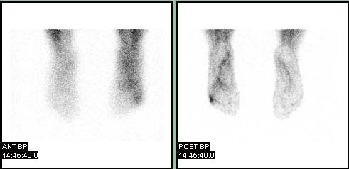
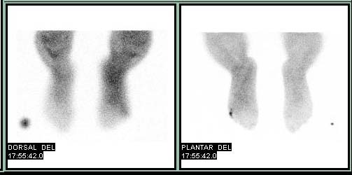
- Radionuclide imaging can be valuable in suspected bone infection, especially early in the course of infection and if multiple foci are suspected or an unusual site is suspected, as in the pelvis.
- Technetium-99 methylene diphosphonate (TC 99m), accumulates in areas of increased bone turnover and is the preferred agent for radionuclide bone imaging (3 phase scan).
- Inflammation and increased vascularity in osteomyelitis result in increased concentration on 99m Tc especially on the 3rd phase (4-6hr later).[1]
The following video and images show bone scans of patients with osteomyelitis.
{{#ev:youtube|X2ShDUfeso0}}
References
- ↑ Schauwecker DS (1992). "The scintigraphic diagnosis of osteomyelitis". AJR Am J Roentgenol. 158 (1): 9–18. doi:10.2214/ajr.158.1.1727365. PMID 1727365.

