Neck of femur fracture CT
Template:Neck of femur Editor-In-Chief: C. Michael Gibson, M.S., M.D. [1]; Associate Editor(s)-in-Chief: Rohan A. Bhimani, M.B.B.S., D.N.B., M.Ch.[2]
Overview
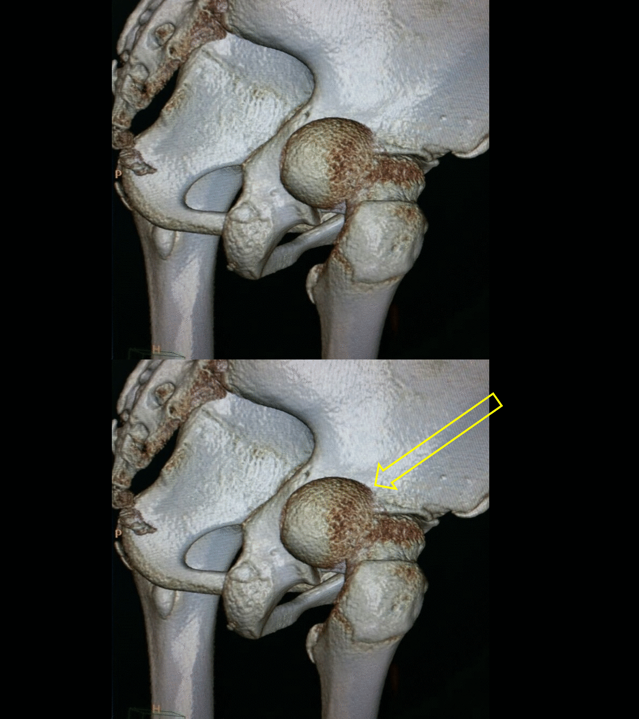
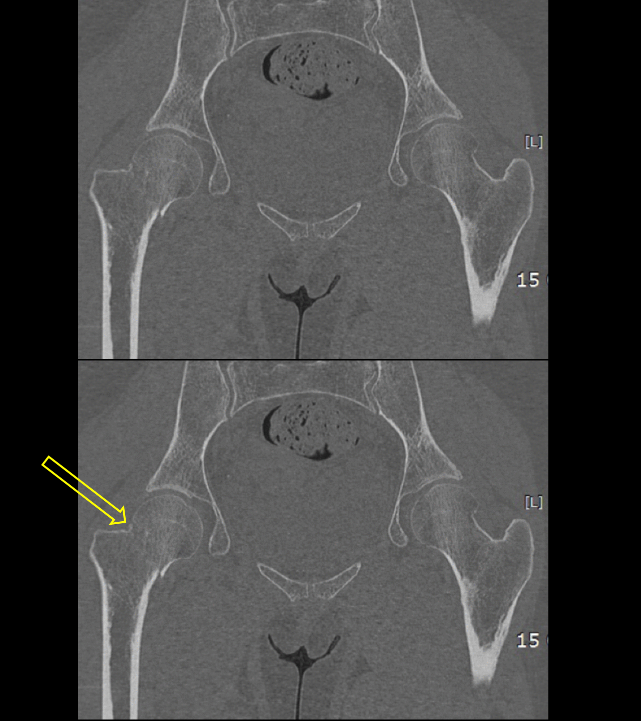
Computed tomography (CT) with two-dimensional reconstruction in the sagittal and coronal planes provides more detailed information than radiographs. CT also helps in fracture fragment orientation and surgical planning.
CT scan
- Computed tomography (CT) with two-dimensional reconstruction in the sagittal and coronal planes provides more detailed information than radiographs.[1][2][3][4]
- CT confirms x ray findings.
- In addition, CT also helps in fracture fragment orientation and surgical planning.
 |
 |
 |
 |
References
- ↑ Tang ZH, Yeoh CS, Tan GM (2017). "Radiographic study of the proximal femur morphology of elderly patients with femoral neck fractures: is there a difference among ethnic groups?". Singapore Med J. 58 (12): 717–720. doi:10.11622/smedj.2016148. PMC 5917059. PMID 27570869.
- ↑ Tiwari S, De Rover WS, Dawson S, Moran C, Sahota O (2015). "Rapid access imaging for occult fractured neck of femur". Osteoporos Int. 26 (1): 407–10. doi:10.1007/s00198-014-2861-8. PMID 25146093.
- ↑ Rockwood, Charles (2010). Rockwood and Green's fractures in adults. Philadelphia, PA: Wolters Kluwer Health/Lippincott Williams & Wilkins. ISBN 9781605476773.
- ↑ Azar, Frederick (2017). Campbell's operative orthopaedics. Philadelphia, PA: Elsevier. ISBN 9780323374620.