Myxoma pathophysiology: Difference between revisions
No edit summary |
No edit summary |
||
| Line 1: | Line 1: | ||
__NOTOC__ | __NOTOC__ | ||
{{Myxoma}} | {{Myxoma}} | ||
{{CMG}} | {{CMG}} {{CZ}} {{AE}} {{AAM}}{{MV}} | ||
==Overview== | ==Overview== | ||
On [[gross pathology]], a gelatinous, irregular surface that fills the [[left atrium]] is characteristic finding of myxoma. On microscopic histopathological analysis, Gamna Bodies consisting of [[fibrosis]] and deposition of [[pigments|iron pigments]] are characteristic findings of myxoma. | On [[gross pathology]], a gelatinous, irregular surface that fills the [[left atrium]] is characteristic finding of myxoma. On microscopic histopathological analysis, Gamna Bodies consisting of [[fibrosis]] and deposition of [[pigments|iron pigments]] are characteristic findings of myxoma. | ||
Revision as of 19:40, 13 November 2015
|
Myxoma Microchapters |
|
Diagnosis |
|---|
|
Treatment |
|
Case Studies |
|
Myxoma pathophysiology On the Web |
|
American Roentgen Ray Society Images of Myxoma pathophysiology |
|
Risk calculators and risk factors for Myxoma pathophysiology |
Editor-In-Chief: C. Michael Gibson, M.S., M.D. [1] Cafer Zorkun, M.D., Ph.D. [2] Associate Editor(s)-in-Chief: Ahmad Al Maradni, M.D. [3]Maria Fernanda Villarreal, M.D. [4]
Overview
On gross pathology, a gelatinous, irregular surface that fills the left atrium is characteristic finding of myxoma. On microscopic histopathological analysis, Gamna Bodies consisting of fibrosis and deposition of iron pigments are characteristic findings of myxoma.
Pathophysiology
Some symptoms of myxoma may be associated with the release of interleukin 6 (IL-6) by the myxoma.[1][2]
Gross Pathology
-
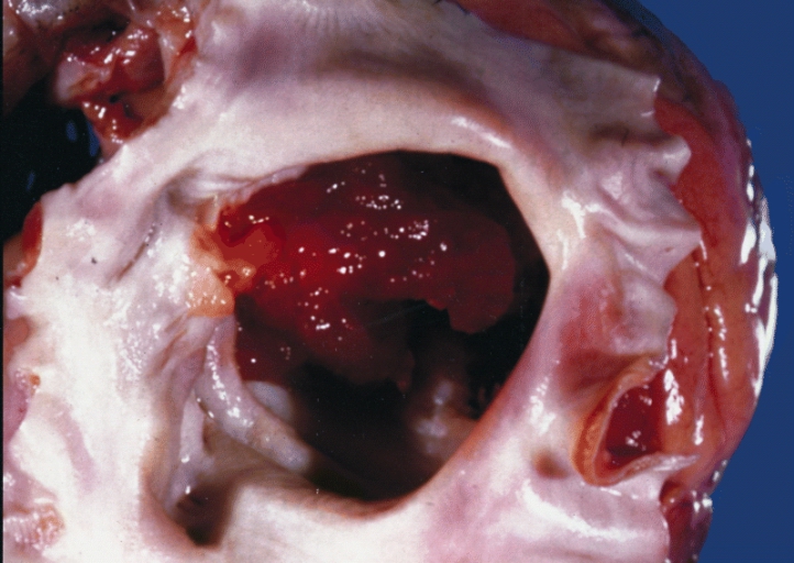
A gelatinous tumor is attached by a narrow pedicle to the atrial septum. The myxoma has an irregular surface and nearly fills the left atrium.
-
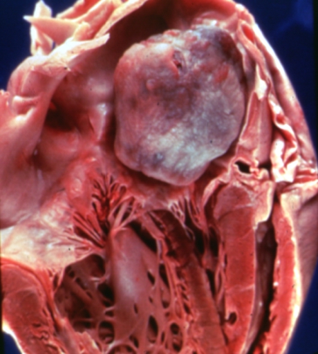
Left Atrial Myxoma


Microscopic Pathology
-
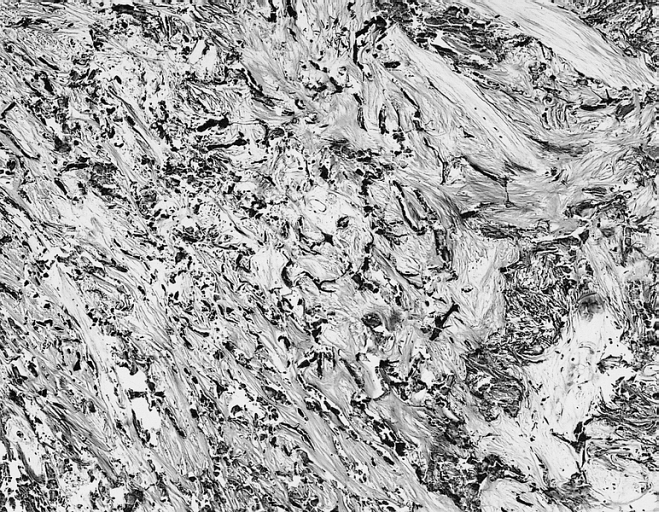
Cardiac Myxoma: Gamna Bodies: A peculiar form of fibrosis with deposition of iron pigment, identical to that seen in the spleens of patients with sickle cell anemia, is not uncommon in myxoma.

-
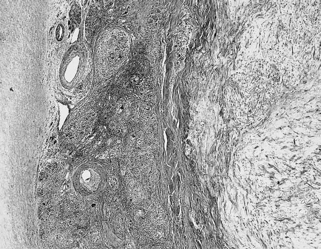
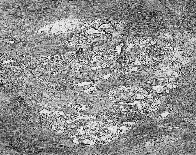
Cardiac Myxoma Common features at the interface with the atrial septum include lymphoid aggregates, smooth muscle bundles, and thick walled vessels which angiographically may look like neovascularization.
-
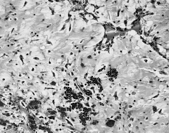
Cardiac Myxoma The extramedullary hematopoiesis seen here is present in about 7 percent of cardiac myxomas.


-
Cardiac Myxoma Glandular structures are seen in less than 5 percent of cases. In this example, they were limited to the base of the myxoma

References
- ↑ Seino Y, Ikeda U, Shimada K. (1993). "Increased expression of interleukin 6 mRNA in cardiac myxomas". Br Heart J. 69 (6): 565–7. PMID 8343326.
- ↑ Jourdan M, Bataille R, Seguin J, Zhang XG, Chaptal PA, Klein B (1990). "Constitutive production of interleukin-6 and immunologic features in cardiac myxomas". Arthritis Rheum. 33 (3): 398–402. PMID 1690543.