Mononucleosis pathophysiology: Difference between revisions
No edit summary |
|||
| Line 1: | Line 1: | ||
{{Mononucleosis}} | {{Mononucleosis}} | ||
{{CMG}} | {{CMG}}; {{AOEIC}} {{LG}} | ||
==Overview== | ==Overview== | ||
Transmission of the [[EBV]] through the air or blood does not normally occur. The incubation period, or the time from infection to appearance of symptoms, ranges from 4 to 6 weeks. Persons with infectious mononucleosis may be able to spread the infection to others for a period of weeks. However, no special precautions or isolation procedures are recommended, since the virus is also found frequently in the saliva of healthy people. In fact, many healthy people can carry and spread the virus intermittently for life. These people are usually the primary reservoir for person-to-person transmission. For this reason, transmission of the virus is almost impossible to prevent. | |||
==Pathophysiology== | |||
*Epstein-Barr virus, frequently referred to as [[EBV]], is a member of the herpesvirus family and one of the most common human viruses. | |||
*Most individuals exposed to people with infectious mononucleosis have previously been infected with EBV and are not at risk for infectious mononucleosis. In addition, transmission of EBV requires intimate contact with the saliva of an infected person. | |||
==Transmission== | ==Transmission== | ||
* Saliva | *'''''Saliva''''' | ||
:* Epstein-Barr virus (EBV) shed for up to 18 months after primary infection | |||
:* Intermittent viral shedding thereafter in asymptomatic sero+ patients | |||
:* Increased viral shedding in immunocompromised patients | |||
* Blood transfusion (rare) | |||
*'''''Blood transfusion''''' (rare) | |||
*Individuals in close living arrangements nearly always pass the infection onto each other, although symptoms may not present for months or even years. | |||
*As with many viral infections, such as [[chicken pox]], antibodies are developed by individuals who become infected with the disease and recover. In most individuals, these [[antibody|antibodies]] remain in their system, creating lifelong immunity to further infections.<ref>{{cite web |title=Mononucleosis -- Causes |url=http://www.emedicinehealth.com/mononucleosis/page2_em.htm |date=12/7/2007 |publisher=eMedicineHealth |accessdate=2008-03-01}}</ref> | |||
==Electron Microscopy== | |||
[[image:Epstein Barr Virus virions EM 10.1371 journal.pbio.0030430.g001-L.JPG|Two Epstein-Barr [[Virion#Structure|virions]]|250px|left]] | |||
<br clear="left"/> | |||
==Microscopic pathology== | |||
Images shown below are courtesy of Professor Peter Anderson DVM PhD and published with permission. [http://www.peir.net © PEIR, University of Alabama at Birmingham, Department of Pathology] | |||
<div align="left"> | |||
<gallery heights="175" widths="175"> | |||
Image:IM 1.jpg|BONE MARROW: INFECTION-ASSOCIATED HEMOPHAGOCYTIC SYNDROME A bone marrow aspirate smear from a child with infection-associated hemophagocytic syndrome secondary to an Epstein-Barr virus infection. On this field there are several large histiocytes. Phagocytosis of nucleated red blood cells, neutrophils, and platelets is evident. The histiocytes have the appearance of reactive cells and should be readily distinguishable from neoplastic histiocytes. (Wright-Giemsa stain) | |||
Image:IM 2.jpg|LOWER RESPIRATORY TRACT: (Supplement) AIL/LYG CD20 staining (for B cells) in the viable tissue shows positive staining of scattered large lymphoid cells (C) which were also the cells that were positive for Epstein-Barr virus by in situ hybridization | |||
Image:IM 3.jpg|LOWER RESPIRATORY TRACT: (Supplement) AIL/LYG CD20 staining (for B cells) in the viable tissue shows positive staining of scattered large lymphoid cells (C) which were also the cells that were positive for Epstein-Barr virus by in situ hybridization | |||
</gallery> | |||
</div> | |||
<div align="left"> | |||
<gallery heights="175" widths="175"> | |||
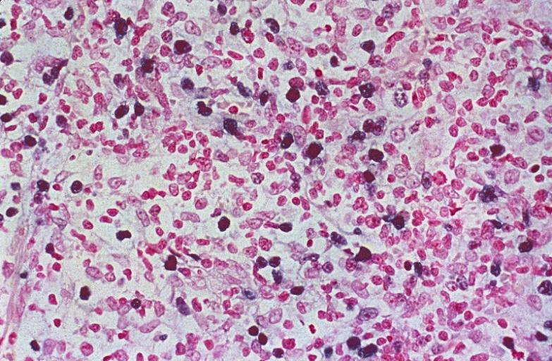
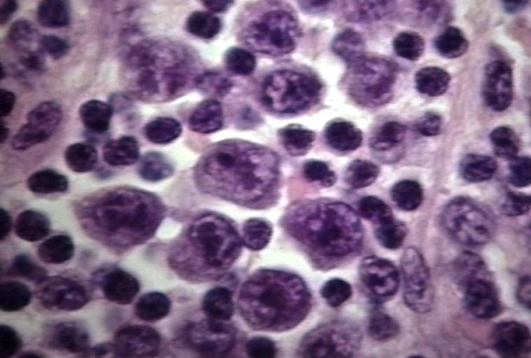
Image:IM 4.jpg|LYMPH NODES-SPLEEN: EPSTEIN-BARR VIRUS-ASSOCIATED INFECTIOUS MONONUCLEOSIS A heterogeneous population is present, including cells mimicking Hodgkin cells. However, the spectrum of cell types and the extensive apoptosis present would not be found in Hodgkin's disease. | |||
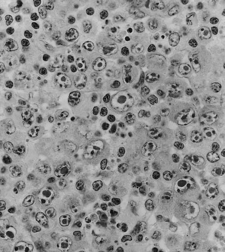
Image:IM 5.jpg|LYMPH NODES-SPLEEN: METASTATIC NASOPHARYNGEAL CARCINOMA IN LYMPH NODE PRESENTING AS METASTATIC CARCINOMA OF UNKNOWN PRIMARY Nuclear labeling of the carcinoma cells for Epstein-Barr virus-encoded RNA strongly suggests that the primary is nasopharyngeal. | |||
Image:IM 6.jpg|BONE MARROW: REACTIVE (ATYPICAL) LYMPHOCYTES This blood smear is from a 19-year-old male college student with infectious mononucleosis. The two reactive (atypical) lymphocytes are large, with abundant cytoplasm and coarse nuclear chromatin, and lack a nucleolus. Cytoplasmic basophilia is radial in distribution and accentuated at the cell margin. In contrast, lymphoblasts are generally smaller, have less cytoplasm with uniform basophilia and more dispersed nuclear chromatin, and may contain a nucleolus. (Wright-Giemsa stain) | |||
</gallery> | |||
</div> | |||
<div align="left"> | |||
<gallery heights="175" widths="175"> | |||
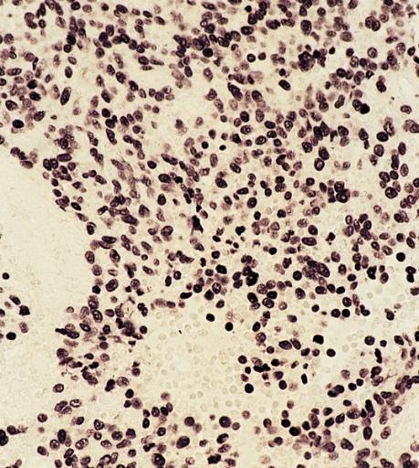
Image:IM 7.jpg|LYMPH NODE: INFECTIOUS MONONUCLEOSIS, LYMPH NODE | |||
Image:IM 8.jpg|BLOOD: INFECTIOUS MONONUCLEOSIS; PERIPHERAL BLOOD | |||
Image:IM 9.jpg|EYE AND OCULAR ADNEXA: BURKITT LYMPHOMA Bilateral involvement. | |||
Image:IM 10.jpg|EYE AND OCULAR ADNEXA: BURKITT LYMPHOMA Lymphoblastic tumor with numerous mitotic figures. | |||
</gallery> | |||
</div> | |||
==References== | ==References== | ||
Revision as of 03:51, 27 February 2012
|
Mononucleosis Microchapters |
|
Diagnosis |
|---|
|
Treatment |
|
Case Studies |
|
Mononucleosis pathophysiology On the Web |
|
American Roentgen Ray Society Images of Mononucleosis pathophysiology |
|
Risk calculators and risk factors for Mononucleosis pathophysiology |
Editor-In-Chief: C. Michael Gibson, M.S., M.D. [1]; Associate Editor(s)-In-Chief: Lakshmi Gopalakrishnan, M.B.B.S. [2]
Overview
Transmission of the EBV through the air or blood does not normally occur. The incubation period, or the time from infection to appearance of symptoms, ranges from 4 to 6 weeks. Persons with infectious mononucleosis may be able to spread the infection to others for a period of weeks. However, no special precautions or isolation procedures are recommended, since the virus is also found frequently in the saliva of healthy people. In fact, many healthy people can carry and spread the virus intermittently for life. These people are usually the primary reservoir for person-to-person transmission. For this reason, transmission of the virus is almost impossible to prevent.
Pathophysiology
- Epstein-Barr virus, frequently referred to as EBV, is a member of the herpesvirus family and one of the most common human viruses.
- Most individuals exposed to people with infectious mononucleosis have previously been infected with EBV and are not at risk for infectious mononucleosis. In addition, transmission of EBV requires intimate contact with the saliva of an infected person.
Transmission
- Saliva
- Epstein-Barr virus (EBV) shed for up to 18 months after primary infection
- Intermittent viral shedding thereafter in asymptomatic sero+ patients
- Increased viral shedding in immunocompromised patients
- Blood transfusion (rare)
- Individuals in close living arrangements nearly always pass the infection onto each other, although symptoms may not present for months or even years.
- As with many viral infections, such as chicken pox, antibodies are developed by individuals who become infected with the disease and recover. In most individuals, these antibodies remain in their system, creating lifelong immunity to further infections.[1]
Electron Microscopy

Microscopic pathology
Images shown below are courtesy of Professor Peter Anderson DVM PhD and published with permission. © PEIR, University of Alabama at Birmingham, Department of Pathology
-
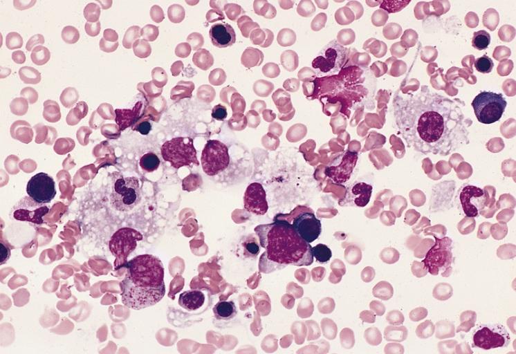
BONE MARROW: INFECTION-ASSOCIATED HEMOPHAGOCYTIC SYNDROME A bone marrow aspirate smear from a child with infection-associated hemophagocytic syndrome secondary to an Epstein-Barr virus infection. On this field there are several large histiocytes. Phagocytosis of nucleated red blood cells, neutrophils, and platelets is evident. The histiocytes have the appearance of reactive cells and should be readily distinguishable from neoplastic histiocytes. (Wright-Giemsa stain)
-
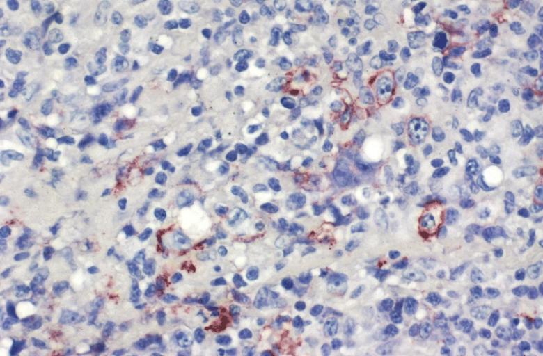
LOWER RESPIRATORY TRACT: (Supplement) AIL/LYG CD20 staining (for B cells) in the viable tissue shows positive staining of scattered large lymphoid cells (C) which were also the cells that were positive for Epstein-Barr virus by in situ hybridization
-
LOWER RESPIRATORY TRACT: (Supplement) AIL/LYG CD20 staining (for B cells) in the viable tissue shows positive staining of scattered large lymphoid cells (C) which were also the cells that were positive for Epstein-Barr virus by in situ hybridization


-
LYMPH NODES-SPLEEN: EPSTEIN-BARR VIRUS-ASSOCIATED INFECTIOUS MONONUCLEOSIS A heterogeneous population is present, including cells mimicking Hodgkin cells. However, the spectrum of cell types and the extensive apoptosis present would not be found in Hodgkin's disease.
-
LYMPH NODES-SPLEEN: METASTATIC NASOPHARYNGEAL CARCINOMA IN LYMPH NODE PRESENTING AS METASTATIC CARCINOMA OF UNKNOWN PRIMARY Nuclear labeling of the carcinoma cells for Epstein-Barr virus-encoded RNA strongly suggests that the primary is nasopharyngeal.
-
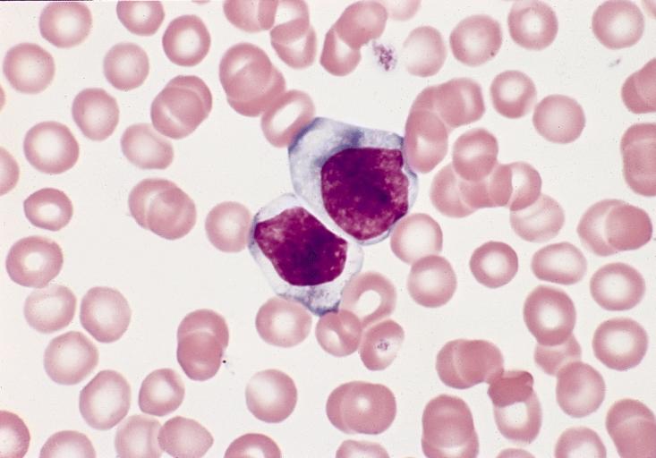
BONE MARROW: REACTIVE (ATYPICAL) LYMPHOCYTES This blood smear is from a 19-year-old male college student with infectious mononucleosis. The two reactive (atypical) lymphocytes are large, with abundant cytoplasm and coarse nuclear chromatin, and lack a nucleolus. Cytoplasmic basophilia is radial in distribution and accentuated at the cell margin. In contrast, lymphoblasts are generally smaller, have less cytoplasm with uniform basophilia and more dispersed nuclear chromatin, and may contain a nucleolus. (Wright-Giemsa stain)


-
LYMPH NODE: INFECTIOUS MONONUCLEOSIS, LYMPH NODE
-
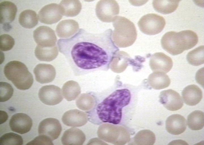
BLOOD: INFECTIOUS MONONUCLEOSIS; PERIPHERAL BLOOD
-
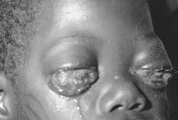
EYE AND OCULAR ADNEXA: BURKITT LYMPHOMA Bilateral involvement.
-

EYE AND OCULAR ADNEXA: BURKITT LYMPHOMA Lymphoblastic tumor with numerous mitotic figures.


References
- ↑ "Mononucleosis -- Causes". eMedicineHealth. 12/7/2007. Retrieved 2008-03-01. Check date values in:
|date=(help)