Medullary thyroid cancer pathophysiology: Difference between revisions
| Line 64: | Line 64: | ||
(2015). http://librepathology.org/wiki/index.php/Medullary_thyroid_carcinoma Accessed on November 12, 2015</ref> | (2015). http://librepathology.org/wiki/index.php/Medullary_thyroid_carcinoma Accessed on November 12, 2015</ref> | ||
Image:Thyroid MedullaryCarcinoma Comedonecrosis LP2 CTR.jpg| Medullary thyroid carcinoma comedonecrosis<ref> Medullary thyroid cancer librepathology | Image:Thyroid MedullaryCarcinoma Comedonecrosis LP2 CTR.jpg| Medullary thyroid carcinoma comedonecrosis<ref> Medullary thyroid cancer librepathology | ||
(2015). http://librepathology.org/wiki/index.php/Medullary_thyroid_carcinoma Accessed on November 12, 2015</ref> | |||
Image:Thyroid MedullaryCarcinoma Comedonecrosis HP CTR.jpg| Medullary thyroid carcinoma comedonecrosis<ref> Medullary thyroid cancer librepathology | |||
(2015). http://librepathology.org/wiki/index.php/Medullary_thyroid_carcinoma Accessed on November 12, 2015</ref> | (2015). http://librepathology.org/wiki/index.php/Medullary_thyroid_carcinoma Accessed on November 12, 2015</ref> | ||
</gallery> | </gallery> | ||
Revision as of 20:52, 12 November 2015
|
Medullary thyroid cancer Microchapters |
|
Differentiating Medullary thyroid cancer from other Diseases |
|---|
|
Diagnosis |
|
Treatment |
|
Case Studies |
|
Medullary thyroid cancer pathophysiology On the Web |
|
American Roentgen Ray Society Images of Medullary thyroid cancer pathophysiology |
|
Risk calculators and risk factors for Medullary thyroid cancer pathophysiology |
Editor-In-Chief: C. Michael Gibson, M.S., M.D. [1]; Associate Editor(s)-in-Chief: Ammu Susheela, M.D. [2]
Overview
Pathogenesis
- Medullary thyroid carcinoma (MTC) is a subtype of thyroid cancer which accounts for 5-10% of all thyroid malignancies 1. It occurs both sporadically (80%) and as a familial form.
- It is characterised by consistent production of a hormonal marker, calcitonin, calcification of both primary and metastatic sites, and association with other endocrine neoplasms. Metastatic involvement may be seen in up to 50% at the time of presentation
- Approximately 25% of reported cases of MTC are familial. Familial MTC syndromes include MEN 2A, which is the most common; MEN 2B; and familial non-MEN syndromes. MTC can secrete calcitonin and other peptide substances.
- Medullary thyroid cancer (MTC) is a form of thyroid carcinoma which originates from the parafollicular cells (C cells), which produce the hormone calcitonin.[1]
Medullary tumors are the third most common of all thyroid cancers. They make up about 3% of all thyroid cancer cases.
Approximately 25% of medullary thyroid cancer is genetic in nature, caused by a mutation in the RET proto-oncogene. This form is classified as familial MTC. When MTC occurs by itself it is termed sporadic MTC. When it coexists with tumors of the parathyroid gland and medullary component of the adrenal glands (pheochromocytoma) it is called multiple endocrine neoplasia type 2 (MEN2).
Genetics
Mutations (DNA changes) in the RET proto-oncogene, located on chromosome 10, lead to the expression of a mutated receptor tyrosine kinase protein, termed RET (REarranged during Transfection). RET is involved in the regulation of cell growth and development and its germline mutation is responsible for nearly all cases of hereditary or familial medullary thyroid carcinoma. Its germline mutation may also be responsible for the development of hyperparathyroidism and pheochromocytoma. Hereditary medullary thyroid cancer is inherited as an autosomal dominant trait, meaning that each child of an affected parent has a 50% probability of inheriting the mutant RET proto-oncogene from the affected parent. DNA analysis makes it possible to identify children who carry the mutant gene; surgical removal of the thyroid in children who carry the mutant gene is curative if the entire thyroid gland is removed at an early age, before there is spread of the tumor. The parathyroid tumors and pheochromocytomas are removed when they cause clinical symptomatology. Hereditary medullary thyroid carcinoma or multiple endocrine neoplasia (MEN2) accounts for approximately 25% of all medullary thyroid carcinomas.
Seventy-five percent of medullary thyroid carcinoma occurs in individuals without an identifiable family history and is assigned the term "sporadic". Individuals who develop sporadic medullary thyroid carcinoma tend to be older and have more extensive disease at the time of initial presentation than those with a family history (screening is likely to be initiated at an early age in the hereditary form). Approximately 25-60% of sporadic medullary thyroid carcinomas have a somatic mutation (one that occurs within a single "parafollicular" cell) of the RET proto-oncogene. This mutation is presumed to be the initiating event, although there could be other as yet unidentified causes.
Associated Conditions
- von Hippel Lindau diseas
- Neurofibromatosis type 1
Gross Pathology
- Medullary thyroid cancer is usually is well circumscribed, gray, white, or yellow in color mass which is gritty to firm consistency.
Microscopic Pathology
- Microscopic features of medullary thyroid cancer is as follows:
Cytoplasm and Nuclei
- Nested with delicate vascular septa
- Trabecular
- Tubular/glandular, pseudo-papillary cells
- Polygonal to spindle to small cells
- Amphophilic, somewhat granular cytoplasm
- Interstitial edema.
- Stroma may have +/-Amyloid deposits with fluffy appearing acellular eosinophilic material in the cytoplasm.
- Stroma is vascular and can show hemorrhage, hyalinised collagen, oedema or metaplastic bone
- Coarse calcification and psammoma bodies may be present
- Nuclei with "neuroendocrine features".
- Small, round nuclei.
- Coarse chromatin (salt and pepper nuclei).
Surrounding Thyroid
- +/-C-cell hyperplasia - seen with familial forms of MTC.
- C cells (AKA parafollicular cell): abundant cytoplasm - clear/pale.
-
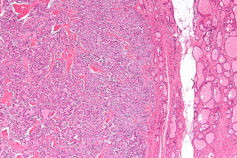
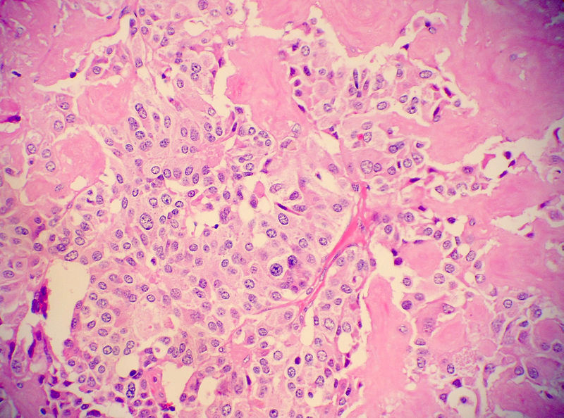
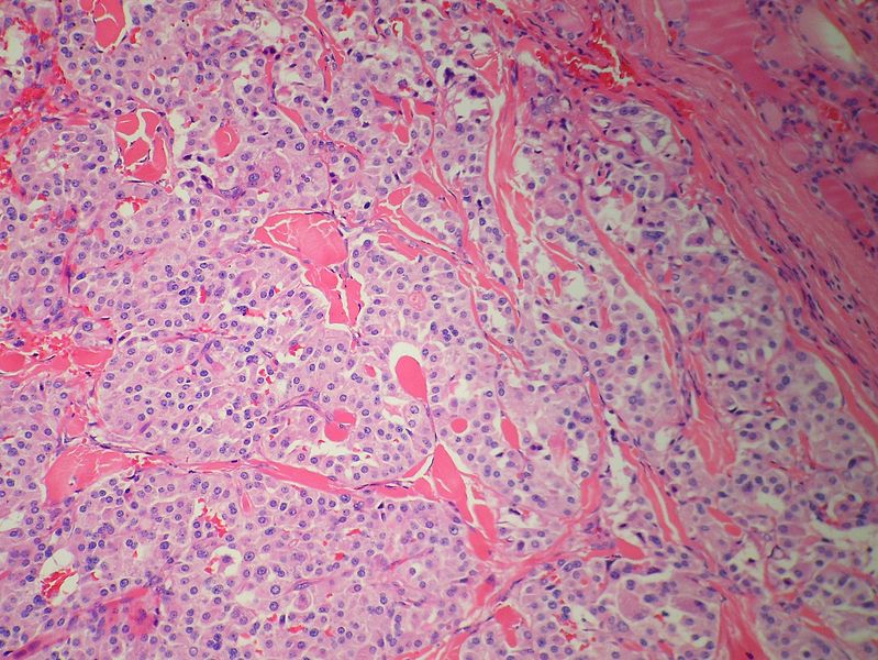
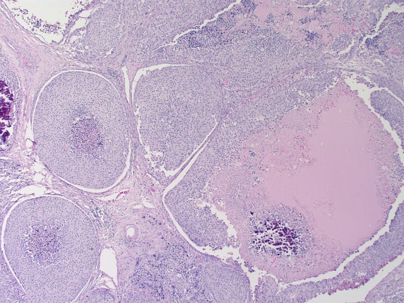
Low magnification micrograph of medullary thyroid carcinoma<ref> Medullary thyroid cancer librepathology
-
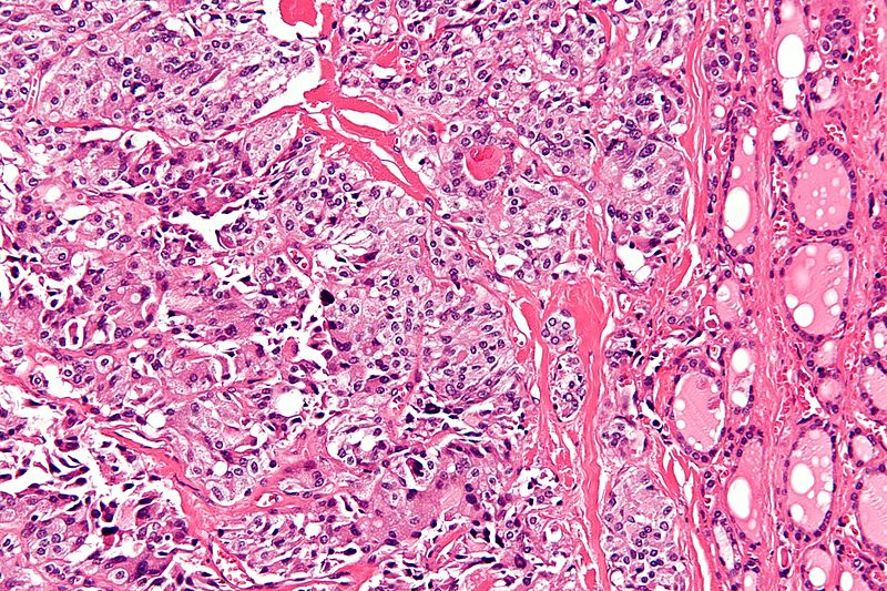
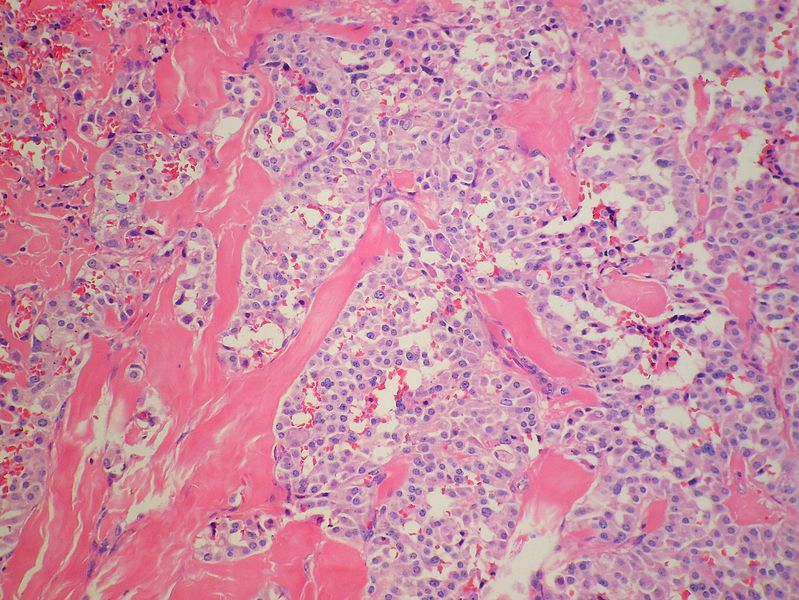
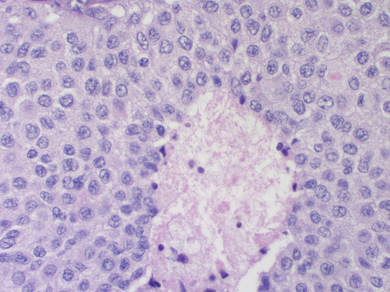
High magnification micrograph of medullary thyroid carcinoma<ref> Medullary thyroid cancer librepathology
-
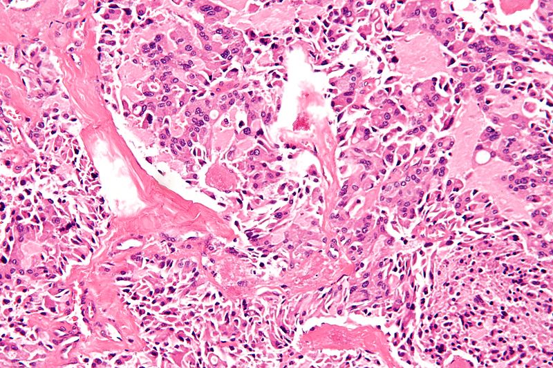
High magnification micrograph of medullary thyroid carcinoma<ref> Medullary thyroid cancer librepathology
-
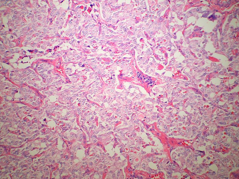
Low magnification micrograph of medullary thyroid carcinoma<ref> Medullary thyroid cancer librepathology
-
Medullary thyroid carcinoma amyloid<ref> Medullary thyroid cancer librepathology
-
Medullary thyroid carcinoma amyloid<ref> Medullary thyroid cancer librepathology
-
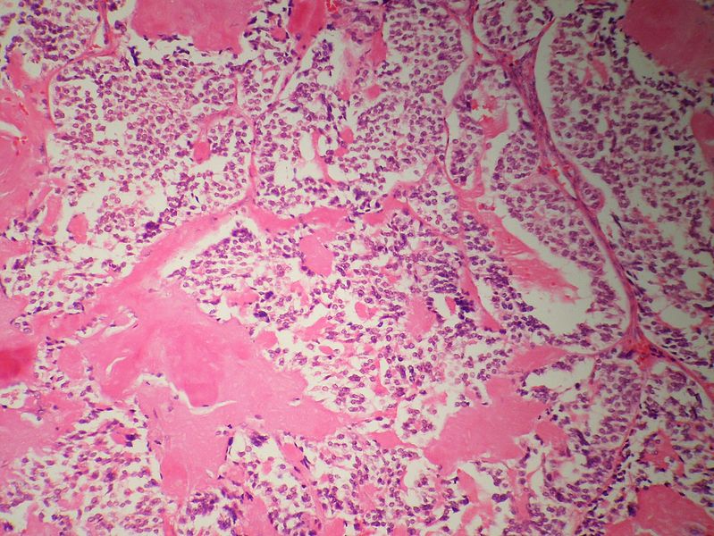
Medullary thyroid carcinoma amyloid<ref> Medullary thyroid cancer librepathology
-
Medullary thyroid carcinoma amyloid<ref> Medullary thyroid cancer librepathology
-
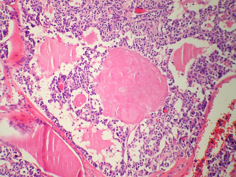
Medullary thyroid carcinoma amyloid<ref> Medullary thyroid cancer librepathology
-
Medullary thyroid carcinoma comedonecrosis<ref> Medullary thyroid cancer librepathology
-
Medullary thyroid carcinoma comedonecrosis<ref> Medullary thyroid cancer librepathology


References
- ↑ Hu MI, Vassilopoulou-Sellin R, Lustig R, Lamont JP. "Thyroid and Parathyroid Cancers" in Pazdur R, Wagman LD, Camphausen KA, Hoskins WJ (Eds) Cancer Management: A Multidisciplinary Approach. 11 ed. 2008.