Meckel's diverticulum laboratory findings
|
Meckel's diverticulum Microchapters |
|
Diagnosis |
|---|
|
Treatment |
|
Case Studies |
|
Meckel's diverticulum laboratory findings On the Web |
|
American Roentgen Ray Society Images of Meckel's diverticulum laboratory findings |
|
Risk calculators and risk factors for Meckel's diverticulum laboratory findings |
Editor-In-Chief: C. Michael Gibson, M.S., M.D. [1]
Please help WikiDoc by adding more content here. It's easy! Click here to learn about editing.
Diagnosis
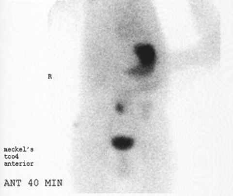
A technetium-99m (99mTc) pertechnetate scan is the investigation of choice to diagnose Meckel's diverticula. This scan detects gastric mucosa; since approximately 50% of symptomatic Meckel's diverticula have ectopic gastric (stomach) cells contained within them, this is displayed as a spot on the scan distant from the stomach itself. Patients with these misplaced gastric cells may experience peptic ulcers as a consequence. Other tests such as colonoscopy and screenings for bleeding disorders should be performed, and angiography can assist in determining the location and severity of bleeding.
Positive Technetium-99m pertechnetate scan
Imaging Findings
- Meckel diverticulum is identified as a saccular, blind-ending structure located on the antimesenteric border of the ileum.
- Meckel diverticulum is usually found in the right lower quadrant and pelvic region.
- The junction of the diverticulum with the ileum may show a mucosal triangular plateau or triradiate fold pattern (represents the site of omphalomesenteric duct attachment to the ileum).
- Filling defects within the diverticulum may represent enteroliths, fecoliths, or foreign bodies.
- Technetium-99m pertechnetate scintigraphy is the modality of choice for evaluating pediatric patients with gastrointestinal hemorrhage and a suspected Meckel diverticulum.
- A Meckel diverticulum containing gastric mucosa will manifest as a small rounded area of increased activity in the right lower quadrant.
- Normal activity will simultaneously appear in the stomach.
