Mast cell leukemia physical examination
|
Mast cell leukemia Microchapters |
|
Diagnosis |
|---|
|
Treatment |
|
Case Studies |
|
Mast cell leukemia physical examination On the Web |
|
American Roentgen Ray Society Images of Mast cell leukemia physical examination |
|
Risk calculators and risk factors for Mast cell leukemia physical examination |
Editor-In-Chief: C. Michael Gibson, M.S., M.D. [1]; Associate Editor(s)-in-Chief: Nawal Muazam M.D.[2]
Overview
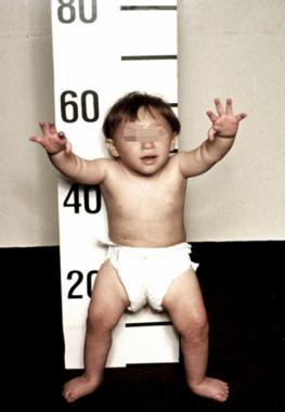
Common physical examination findings of mast cell leukemia include hepatomegaly, splenomegaly, lymphadenopathy, bone abnormalities, and ascites.
Physical Examination
Common physical examination findings of mast cell leukemia include:[1][2]
- Hepatomegaly
- Hypotention
- Splenomegaly
- Lymphadenopathy
- Tachycardia
- Bone abnormalities
- Ascites
- Weight loss
- Asthenia
Physical Examination
Physical examination of patients with [disease name] is usually normal.
OR
Physical examination of patients with [disease name] is usually remarkable for [finding 1], [finding 2], and [finding 3].
OR
The presence of [finding(s)] on physical examination is diagnostic of [disease name].
OR
The presence of [finding(s)] on physical examination is highly suggestive of [disease name].
Appearance of the Patient
- Patients with [disease name] usually appear [general appearance].
Vital Signs
- High-grade / low-grade fever
- Hypothermia / hyperthermia may be present
- Tachycardia with regular pulse or (ir)regularly irregular pulse
- Bradycardia with regular pulse or (ir)regularly irregular pulse
- Tachypnea / bradypnea
- Kussmal respirations may be present in _____ (advanced disease state)
- Weak/bounding pulse / pulsus alternans / paradoxical pulse / asymmetric pulse
- High/low blood pressure with normal pulse pressure / wide pulse pressure / narrow pulse pressure
Skin
- Skin examination of patients with [disease name] is usually normal.
OR
-
Description (Adapted from Dermatology Atlas)
-
Description (Adapted from Dermatology Atlas)

HEENT
- HEENT examination of patients with [disease name] is usually normal.
OR
- Abnormalities of the head/hair may include ___
- Evidence of trauma
- Icteric sclera
- Nystagmus
- Extra-ocular movements may be abnormal
- Pupils non-reactive to light / non-reactive to accommodation / non-reactive to neither light nor accommodation
- Ophthalmoscopic exam may be abnormal with findings of ___
- Hearing acuity may be reduced
- Weber test may be abnormal (Note: A positive Weber test is considered a normal finding / A negative Weber test is considered an abnormal finding. To avoid confusion, you may write "abnormal Weber test".)
- Rinne test may be positive (Note: A positive Rinne test is considered a normal finding / A negative Rinne test is considered an abnormal finding. To avoid confusion, you may write "abnormal Rinne test".)
- Exudate from the ear canal
- Tenderness upon palpation of the ear pinnae/tragus (anterior to ear canal)
- Inflamed nares / congested nares
- Purulent exudate from the nares
- Facial tenderness
- Erythematous throat with/without tonsillar swelling, exudates, and/or petechiae
Neck
- Neck examination of patients with [disease name] is usually normal.
OR
- Jugular venous distension
- Carotid bruits may be auscultated unilaterally/bilaterally using the bell/diaphragm of the otoscope
- Lymphadenopathy (describe location, size, tenderness, mobility, and symmetry)
- Thyromegaly / thyroid nodules
- Hepatojugular reflux
Lungs
- Pulmonary examination of patients with [disease name] is usually normal.
OR
- Asymmetric chest expansion OR decreased chest expansion
- Lungs are hyporesonant OR hyperresonant
- Fine/coarse crackles upon auscultation of the lung bases/apices unilaterally/bilaterally
- Rhonchi
- Vesicular breath sounds OR distant breath sounds
- Expiratory wheezing OR inspiratory wheezing with normal OR delayed expiratory phase
- Wheezing may be present
- Egophony present/absent
- Bronchophony present/absent
- Normal/reduced tactile fremitus
Heart
- Cardiovascular examination of patients with [disease name] is usually normal.
OR
- Chest tenderness upon palpation
- PMI within 2 cm of the sternum (PMI) / Displaced point of maximal impulse (PMI) suggestive of ____
- Heave / thrill
- Friction rub
- S1
- S2
- S3
- S4
- Gallops
- A high/low grade early/late systolic murmur / diastolic murmur best heard at the base/apex/(specific valve region) may be heard using the bell/diaphgram of the stethoscope
Abdomen
- Abdominal examination of patients with [disease name] is usually normal.
OR
- Abdominal distention
- Abdominal tenderness in the right/left upper/lower abdominal quadrant
- Rebound tenderness (positive Blumberg sign)
- A palpable abdominal mass in the right/left upper/lower abdominal quadrant
- Guarding may be present
- Hepatomegaly / splenomegaly / hepatosplenomegaly
- Additional findings, such as obturator test, psoas test, McBurney point test, Murphy test
Back
- Back examination of patients with [disease name] is usually normal.
OR
- Point tenderness over __ vertebrae (e.g. L3-L4)
- Sacral edema
- Costovertebral angle tenderness bilaterally/unilaterally
- Buffalo hump
Genitourinary
- Genitourinary examination of patients with [disease name] is usually normal.
OR
- A pelvic/adnexal mass may be palpated
- Inflamed mucosa
- Clear/(color), foul-smelling/odorless penile/vaginal discharge
Neuromuscular
- Neuromuscular examination of patients with [disease name] is usually normal.
OR
- Patient is usually oriented to persons, place, and time
References
- ↑ Joris, Magalie; Georgin-Lavialle, Sophie; Chandesris, Marie-Olivia; Lhermitte, Ludovic; Claisse, Jean-François; Canioni, Danielle; Hanssens, Katia; Damaj, Gandhi; Hermine, Olivier; Hamidou, Mohammed (2012). "Mast Cell Leukaemia: c-KIT Mutations Are Not Always Positive". Case Reports in Hematology. 2012: 1–6. doi:10.1155/2012/517546. ISSN 2090-6560.
- ↑ Georgin-Lavialle, S.; Lhermitte, L.; Dubreuil, P.; Chandesris, M.-O.; Hermine, O.; Damaj, G. (2012). "Mast cell leukemia". Blood. 121 (8): 1285–1295. doi:10.1182/blood-2012-07-442400. ISSN 0006-4971.