Ischemic stroke CT: Difference between revisions
Aysha Aslam (talk | contribs) No edit summary |
No edit summary |
||
| Line 15: | Line 15: | ||
Image:ACA-infarction-001.jpg|CT | Image:ACA-infarction-001.jpg|CT | ||
Image:ACA-infarction-002.jpg|CT | Image:ACA-infarction-002.jpg|CT | ||
</gallery> | </gallery>'''For AHA/ASA guidelines for CT scan in patients with ischemic stroke, please''' [[AHA/ASA guideline recommendations for of Early management of acute ischemic stroke#Parenchymal Brain Imaging|click here]] | ||
</div> | </div> | ||
Revision as of 14:24, 12 January 2023
|
Ischemic Stroke Microchapters |
|
Diagnosis |
|---|
|
Treatment |
|
Case Studies |
|
Ischemic stroke CT On the Web |
|
American Roentgen Ray Society Images of Ischemic stroke CT |
Editor-In-Chief: C. Michael Gibson, M.S., M.D. [1]Associate Editor(s)-in-Chief: Aysha Anwar, M.B.B.S[2]
Overview
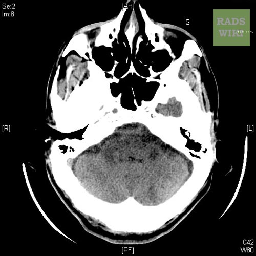
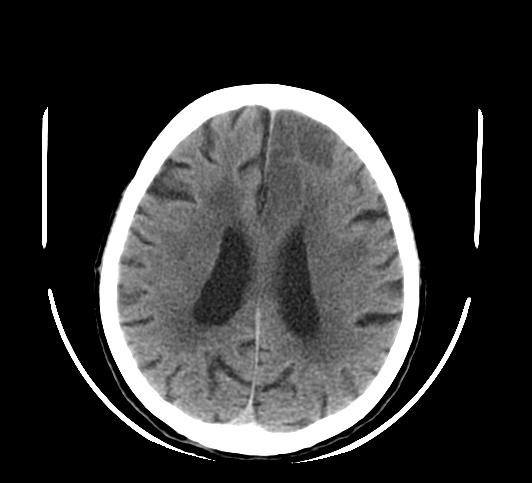
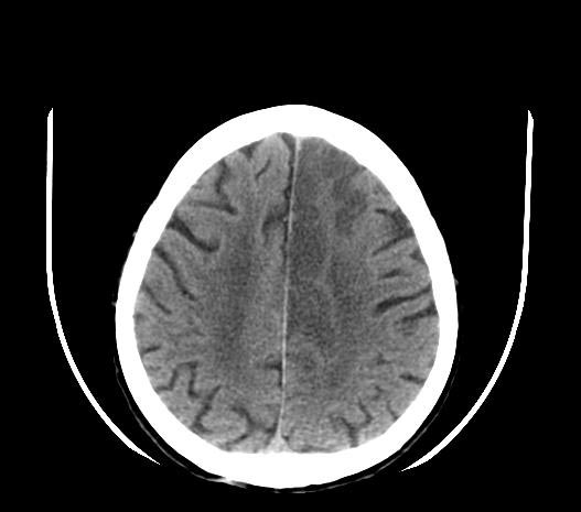
CT scan without contrast is the initial test performed to diagnose ischemic stroke and rule out hemorrhagic stroke. In the emergency setting, the sensitivity and specificity of CT scan without contrast is 16% and 96% respectively [1] The sensitivity and specificity for detection of early ischemia is enhanced with perfusion CT scan.
CT
CT scan without contrast is the initial test performed to diagnose ischemic stroke and rule out hemorrhagic stroke. In the emergency setting, the sensitivity and specificity of CT scan without contrast is 16% and 96% respectively [1] The sensitivity and specificity for detection of early ischemia is enhanced with perfusion CT scan. The main advantages of CT scan compared to MRI includes it being rapid, low cost, availability in acute setting, safe in patients with metallic implants such as pacemakers, implantable defibrillators. Some of the disadvantages which may limit its use include its decreased sensitivity and specificity to detect lacunar and posterior fossa infarcts. CT scan with contrast cannot be used in patients with renal failure.[2]
For AHA/ASA guidelines for CT scan in patients with ischemic stroke, please click here
-
CT
-
CT
-
CT


References
- ↑ 1.0 1.1 Chalela, J (2007). "Magnetic resonance imaging and computed tomography in emergency assessment of patients with suspected acute stroke: a prospective comparison". Lancet. 369 (9558): 293–8. PMID 17258669. Retrieved 2008-01-22. Unknown parameter
|coauthors=ignored (help) - ↑ Wintermark M, Sanelli PC, Albers GW, Bello J, Derdeyn C, Hetts SW; et al. (2013). "Imaging recommendations for acute stroke and transient ischemic attack patients: A joint statement by the American Society of Neuroradiology, the American College of Radiology, and the Society of NeuroInterventional Surgery". AJNR Am J Neuroradiol. 34 (11): E117–27. doi:10.3174/ajnr.A3690. PMC 4072500. PMID 23907247.