Intravenous leiomyomatosis: Difference between revisions
Jump to navigation
Jump to search
No edit summary |
No edit summary |
||
| Line 16: | Line 16: | ||
==Pathophysiology== | ==Pathophysiology== | ||
*The etiology of intravenous leiomyomatosis is unclear. All described patients are female, and most are white, premenopausal, and parous. | *The etiology of intravenous leiomyomatosis is unclear. All described patients are female, and most are white, premenopausal, and parous. | ||
*The patients may be asymptomatic or have symptoms of uterine leiomyomas. | *The patients may be asymptomatic or have symptoms of uterine leiomyomas. | ||
*Patients with obstruction of the right atrium may present with syncopal episodes, dyspnea on exertion, shortness of breath, etc. | *Patients with obstruction of the right atrium may present with syncopal episodes, dyspnea on exertion, shortness of breath, etc. | ||
| Line 24: | Line 23: | ||
*When the intravenous catheter is involved the differential diagnosis should include renal malignancies and primary leiomyoma or sarcoma, as well as thrombosis of the intravenous catheter. | *When the intravenous catheter is involved the differential diagnosis should include renal malignancies and primary leiomyoma or sarcoma, as well as thrombosis of the intravenous catheter. | ||
*Intravenous leiomyomatosis should be considered in young women with cardiac symptoms who have a right atrial mass as well as a pelvic mass or who have previously undergone hysterectomy for leiomyoma uterus with intravenous involvement. | *Intravenous leiomyomatosis should be considered in young women with cardiac symptoms who have a right atrial mass as well as a pelvic mass or who have previously undergone hysterectomy for leiomyoma uterus with intravenous involvement. | ||
==Epidemiology and Demographics== | |||
*The median age is 45 years, with patients ranging from 26 to 70 years old. | |||
== Natural History, Complications and Prognosis== | == Natural History, Complications and Prognosis== | ||
| Line 31: | Line 32: | ||
*Intravenous leiomyomatosis should not be confused with benign metastasizing leiomyoma, in which a benign uterine leiomyoma is associated with a benign smooth muscle tumor located in the parenchyma of a distant organ, such as lung. | *Intravenous leiomyomatosis should not be confused with benign metastasizing leiomyoma, in which a benign uterine leiomyoma is associated with a benign smooth muscle tumor located in the parenchyma of a distant organ, such as lung. | ||
*Intravenous leiomyomatosis is confined to vessels, whereas benign metastasizing leiomyoma shows no relation to vascular channels. | *Intravenous leiomyomatosis is confined to vessels, whereas benign metastasizing leiomyoma shows no relation to vascular channels. | ||
== Diagnosis == | |||
=== Symptoms === | |||
*Symptoms of [disease name] may include the following: | |||
:*[symptom 1] | |||
:*[symptom 2] | |||
:*[symptom 3] | |||
=== Physical Examination === | |||
*Physical examination may be remarkable for: | |||
:*[finding 1] | |||
:*[finding 2] | |||
:*[finding 3] | |||
==Images== | ==Images== | ||
===Example #1=== | ===Example #1=== | ||
Revision as of 20:05, 14 April 2016
Editor-In-Chief: C. Michael Gibson, M.S., M.D. [1]
Associate Editor-In-Chief: Cafer Zorkun, M.D., Ph.D. [2]; Ammu Susheela, M.D. [3]
Synonyms and keywords: Nesidioblastoma
Overview
- Intravenous leiomyomatosis (IVLM) is characterized by the extension into venous channels of histologically benign smooth muscle tumor arising from either the wall of a vessel or from a uterine leiomyoma.
- Fewer than 100 cases have been reported in all, and only 14 cases involved intracardiac extension from the IVC.
- In one reported case, this slowly growing invasive neoplasm extended not only into the heart but into both pulmonary arteries as well. [1]
Pathophysiology
- The etiology of intravenous leiomyomatosis is unclear. All described patients are female, and most are white, premenopausal, and parous.
- The patients may be asymptomatic or have symptoms of uterine leiomyomas.
- Patients with obstruction of the right atrium may present with syncopal episodes, dyspnea on exertion, shortness of breath, etc.
- The tumor is slow growing, and the prognosis is favorable.
Differentiating Intravenous Leiomyomatosis from other Diseases
- When the intravenous catheter is involved the differential diagnosis should include renal malignancies and primary leiomyoma or sarcoma, as well as thrombosis of the intravenous catheter.
- Intravenous leiomyomatosis should be considered in young women with cardiac symptoms who have a right atrial mass as well as a pelvic mass or who have previously undergone hysterectomy for leiomyoma uterus with intravenous involvement.
Epidemiology and Demographics
- The median age is 45 years, with patients ranging from 26 to 70 years old.
Natural History, Complications and Prognosis
- Although embolization of the tumor represents a theoretical risk, this has not been reported.
- The tumor can recur, and repeat operation may be necessary.
- Most reported deaths involved extension of the tumor into the heart, with death due to mechanical obstruction rather than the neoplastic process per se
- Intravenous leiomyomatosis should not be confused with benign metastasizing leiomyoma, in which a benign uterine leiomyoma is associated with a benign smooth muscle tumor located in the parenchyma of a distant organ, such as lung.
- Intravenous leiomyomatosis is confined to vessels, whereas benign metastasizing leiomyoma shows no relation to vascular channels.
Diagnosis
Symptoms
- Symptoms of [disease name] may include the following:
- [symptom 1]
- [symptom 2]
- [symptom 3]
Physical Examination
- Physical examination may be remarkable for:
- [finding 1]
- [finding 2]
- [finding 3]
Images
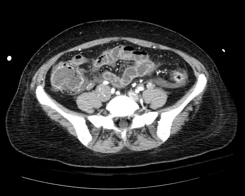
Example #1
Patient presented with S.O.B. one year after hysterectomy for a leiomyomatous uterus.
-
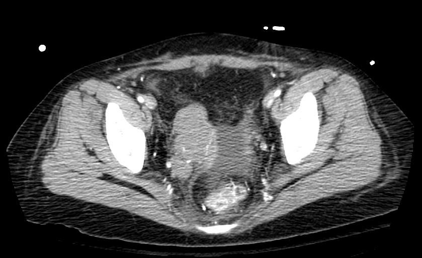
CT in Intravenous leiomyomatosis
-


Related Chapters
- Uterine leiomyoma
- Benign metastasizing leiomyoma
References
- ↑ DJ Kaszar-Seibert, GP Gauvin, PA Rogoff, FJ Vittimberga, S Margolis, AD Hilgenberg, DK Saal, and GO Goldsmith. Intracardiac extension of intravenous leiomyomatosis. Radiology 1988 168: 409-410.