Horner's syndrome
| Horner's syndrome | |
 | |
|---|---|
| Horner's Syndrome |
|
Horner's syndrome Microchapters |
|
Diagnosis |
|---|
|
Treatment |
|
Case Studies |
|
Horner's syndrome On the Web |
|
American Roentgen Ray Society Images of Horner's syndrome |
For patient information, click here
Editor-In-Chief: C. Michael Gibson, M.S., M.D. [1]
Synonyms and keywords: Bernard-Horner syndrome; oculosympathetic palsy
Overview
Horner's syndrome is a clinical syndrome caused by damage to the sympathetic nervous system.
Signs
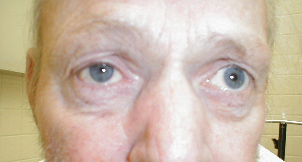
Signs found in all patients on affected side of face include ptosis (drooping upper eyelid from loss of sympathetic innervation to the Müller-Rouget muscle), upside-down ptosis (slight elevation of the lower lid), and miosis (constricted pupil) and dilation lag. Enophthalmos (the impression that the eye is sunk in) and anhidrosis (decreased sweating) on the affected side of the face, loss of ciliospinal reflex and blood shot conjunctiva may occur depending on the site of lesion.
In children Horner's syndrome sometimes leads to a difference in eye color between the two eyes (heterochromia).[1] This happens because a lack of sympathetic stimulation in childhood interferes with melanin pigmentation of the melanocytes in the superficial stroma of the iris.
History
It is named after Johann Friedrich Horner, the Swiss ophthalmologist who first described the syndrome in 1869.[2][3] Several others had previously described cases, but "Horner's syndrome" is most prevalent. In France, Claude Bernard is also eponymised with the condition being called "syndrome Bernard-Horner".
Differential diagnosis of causes of Horner's syndrome
Horner's syndrome is usually acquired but may also be congenital (inborn) or iatrogenic (caused by medical treatment). Although most causes are relatively benign, Horner's syndrome may reflect serious pathology in the neck or chest (such as a Pancoast tumor or thyrocervical venous dilatation) and hence requires workup.
- Due to lesion of one side of the cervical sympathetic chain which affects on the same side of the lesion
- PICA syndrome
- Cluster headache - combination termed Horton's headache[4]
- Trauma - base of neck, usually blunt trauma.
- Middle ear infection
- Tumors - often bronchogenic carcinoma of the superior fissure (Pancoast tumor)
- Thoracic aortic aneurysm
- Neurofibromatosis type 1
- Goitre
- Dissecting aortic aneurysm
- Thyroid carcinoma
- Multiple sclerosis
- Carotid artery dissection
- Klumpke paralysis
- Cavernous sinus thrombosis
- Sympathectomy
- Syringomyelia
- Nerve blocks, such as cervical plexus block, stellate ganglion or interscalene block
- Brainstem stroke
- Carotid body tumor
- Lymphoma
- Mediastinal mass
- Metastasis
- Parotid gland tumor
- Tuberculosis adenitis
Pathophysiology
Horner's syndrome is due to a deficiency of sympathetic activity. The site of lesion to the sympathetic outflow is on the ipsilateral side of the symptoms. The following are examples of conditions that cause the clinical appearance of Horner's syndrome:
- First-order neuron disorder: Central lesions that involve the hypothalamospinal pathway (e.g. transection of the cervical spinal cord).
- Second-order neuron disorder: Preganglionic lesions (e.g. compression of the sympathetic chain by a lung tumor).
- Third-order neuron disorder: Postganglionic lesions at the level of the internal carotid artery (e.g. a tumor in the cavernous sinus).
Diagnosis
Three tests are useful in confirming the presence and severity of Horner's syndrome:
- Cocaine drop test - Cocaine blocks the reuptake of norepinephrine resulting in the dilation of a normal pupil. The pupil will fail to dilate in Horner's syndrome.
- Paredrine test
- Dilation lag test
It is important to distinguish the ptosis caused by Horner's syndrome from the ptosis caused by a lesion to the oculomotor nerve. In the former, the ptosis occurs with a constricted pupil (due to a loss of sympathetics to the eye), whereas in the latter, the ptosis occurs with a dilated pupil (due to a loss of innervation to the sphincter pupillae). In an actual clinical setting, however, these two different ptoses are fairly easy to distinguish. In addition to the blown pupil in a CNIII (oculomotor nerve) lesion, this ptosis is much more severe, occasionally occluding the whole eye. The ptosis of Horner's syndrome can be quite mild or barely noticeable.
-

Horner's Syndrome
-
Horner's Syndrome

Related Chapters
References
- ↑ Gesundheit B, Greenberg M (2005). "Medical mystery: brown eye and blue eye--the answer". N Engl J Med. 353 (22): 2409–10. PMID 16319395.
- ↑ Horner JF. Über eine Form von Ptosis. Klin Monatsbl Augenheilk 1869;7:193-8.
- ↑ Template:WhoNamedIt
- ↑ Graff JM, Lee AG (February 21, 2005). "Horner's Syndrome (due to Cluster Headache): 46 y.o. man presenting with headache and ptosis". Ophthalmology Grand Rounds. The University of Iowa. Retrieved 2006-09-22.
Template:PNS diseases of the nervous system