Hepatocellular carcinoma pathophysiology: Difference between revisions
(Mahshid) |
|||
| Line 1: | Line 1: | ||
<div style="-webkit-user-select: none;"> | <div style="-webkit-user-select: none;"> | ||
{|class="infobox" style="position: fixed; top: 65%; right: 10px; margin: 0 0 0 0; border: 0; float: right; | {| class="infobox" style="position: fixed; top: 65%; right: 10px; margin: 0 0 0 0; border: 0; float: right;" | ||
|- | |- | ||
| {{#ev:youtube|https://https://www.youtube.com/watch?v=zv04qtEM8qw|350}} | | {{#ev:youtube|https://https://www.youtube.com/watch?v=zv04qtEM8qw|350}} | ||
| Line 7: | Line 7: | ||
__NOTOC__ | __NOTOC__ | ||
{{Hepatocellular carcinoma}} | {{Hepatocellular carcinoma}} | ||
{{CMG}}{{AE}}{{PSD}} {{MJK}} | {{CMG}}{{AE}}{{SH}} {{PSD}} {{MJK}} | ||
==Overview== | ==Overview== | ||
| Line 15: | Line 15: | ||
On gross pathology, hepatocellular carcinoma have the following characteristic findings: | On gross pathology, hepatocellular carcinoma have the following characteristic findings: | ||
* Nodular or diffusely infiltrative | * Nodular or diffusely infiltrative. | ||
:* The nodular type may be unifocal (large mass) or multifocal (when developed as a complication of cirrhosis). Tumor nodules are round to oval, grey or green (if the tumor produces bile), well circumscribed but not encapsulated | :* The nodular type may be unifocal (large mass) or multifocal (when developed as a complication of cirrhosis). Tumor nodules are round to oval, grey or green (if the tumor produces bile), well circumscribed but not encapsulated. | ||
:* | :* In about fifty percent of the cases, the tumors are multifocal where as some authors have suggested it to be around 75 percent. | ||
:* The | :* The portal vein is infiltrated by the poorly circumcised diffused type and the hepatic veins are rarely infiltrated. | ||
* Pale in relation to surrounding liver or green (due to bile secretion) | * Pale in relation to surrounding liver or green (due to bile secretion) | ||
<gallery> | <gallery> | ||
| Line 46: | Line 44: | ||
{{WikiDoc Help Menu}} | {{WikiDoc Help Menu}} | ||
{{WikiDoc Sources}} | {{WikiDoc Sources}} | ||
[[Category:Disease]] | [[Category:Disease]] | ||
Revision as of 18:44, 20 December 2017
| https://https://www.youtube.com/watch?v=zv04qtEM8qw%7C350}} |
|
Hepatocellular carcinoma Microchapters |
|
Differentiating Hepatocellular carcinoma from other Diseases |
|---|
|
Diagnosis |
|
Treatment |
|
Case Studies |
|
Hepatocellular carcinoma pathophysiology On the Web |
|
American Roentgen Ray Society Images of Hepatocellular carcinoma pathophysiology |
|
Risk calculators and risk factors for Hepatocellular carcinoma pathophysiology |
Editor-In-Chief: C. Michael Gibson, M.S., M.D. [1]Associate Editor(s)-in-Chief: Dildar Hussain, MBBS [2] Parminder Dhingra, M.D. [3] Mohamad Alkateb, MBBCh [4]
Overview
On microscopic histopathological analysis, large polygonal tumours cells with graunular eosinophilic cytoplasm or layered dense collagen bundles are characteristic findings of hepatocellular carcinoma.
Gross Pathology
On gross pathology, hepatocellular carcinoma have the following characteristic findings:
- Nodular or diffusely infiltrative.
- The nodular type may be unifocal (large mass) or multifocal (when developed as a complication of cirrhosis). Tumor nodules are round to oval, grey or green (if the tumor produces bile), well circumscribed but not encapsulated.
- In about fifty percent of the cases, the tumors are multifocal where as some authors have suggested it to be around 75 percent.
- The portal vein is infiltrated by the poorly circumcised diffused type and the hepatic veins are rarely infiltrated.
- Pale in relation to surrounding liver or green (due to bile secretion)
-
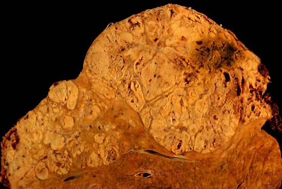
Hepatocelluler carcinoma. The image shows a longitudinal slice taken through the full length of the liver.
(Courtesy of Ed Uthman, MD) -

The tumor is at the top, cirrhotic liver at the bottom, and a fibrous reaction in between. Hepatocellular carcinomas can have a variety of gross patterns, including multinodular / multifocal, such as this one.
(Courtesy of Ed Uthman, MD)


Microscopic Pathology
On microscopic histopathological analysis, hepatocellular carcinoma have the following characteristic findings:
- Large polygonal tumours cells with:
- Graunular eosinophilic cytoplasm
- Low NC ratio
- Layered dense collagen bundles [1]
Videos
{{#ev:youtube|qsR92joabBg}}