Hepatitis D causes: Difference between revisions
Joao Silva (talk | contribs) |
Joao Silva (talk | contribs) |
||
| Line 56: | Line 56: | ||
These elements are only assembled in the presence of the [[hepatitis B virus]] (helper virus). [[HBsAg]] and HDAg-L are necessary and sufficient for virus assembly, whereas [[HDV]] RNA or HDAg-S are not required, but are present, in viral particles. The primary initiation event for [[HDV]] assembly is the interaction of HDAg-L with [[HBsAg]]. HDAg is localized in the [[nuclei]] while and [[HBsAg]] is present in the [[cytoplasm]] of the infected cells. The mechanism of interaction between these two proteins remains unknown. That are different models that try to explain the mechanisms of viral RNA transcription and replication.<ref name=WHO>{{cite web | title = Hepatitis D | url = http://www.who.int/csr/disease/hepatitis/HepatitisD_whocdscsrncs2001_1.pdf }}</ref><ref name="pmid9621000">{{cite journal| author=Modahl LE, Lai MM| title=Transcription of hepatitis delta antigen mRNA continues throughout hepatitis delta virus (HDV) replication: a new model of HDV RNA transcription and replication. | journal=J Virol | year= 1998 | volume= 72 | issue= 7 | pages= 5449-56 | pmid=9621000 | doi= | pmc=PMC110180 | url=http://www.ncbi.nlm.nih.gov/entrez/eutils/elink.fcgi?dbfrom=pubmed&tool=sumsearch.org/cite&retmode=ref&cmd=prlinks&id=9621000 }} </ref><ref>{{cite book | last = Fields | first = Bernard | title = Fields virology | publisher = Wolters Kluwer Health/Lippincott Williams & Wilkins | location = Philadelphia | year = 2013 | isbn = 1451105630 }}</ref> | These elements are only assembled in the presence of the [[hepatitis B virus]] (helper virus). [[HBsAg]] and HDAg-L are necessary and sufficient for virus assembly, whereas [[HDV]] RNA or HDAg-S are not required, but are present, in viral particles. The primary initiation event for [[HDV]] assembly is the interaction of HDAg-L with [[HBsAg]]. HDAg is localized in the [[nuclei]] while and [[HBsAg]] is present in the [[cytoplasm]] of the infected cells. The mechanism of interaction between these two proteins remains unknown. That are different models that try to explain the mechanisms of viral RNA transcription and replication.<ref name=WHO>{{cite web | title = Hepatitis D | url = http://www.who.int/csr/disease/hepatitis/HepatitisD_whocdscsrncs2001_1.pdf }}</ref><ref name="pmid9621000">{{cite journal| author=Modahl LE, Lai MM| title=Transcription of hepatitis delta antigen mRNA continues throughout hepatitis delta virus (HDV) replication: a new model of HDV RNA transcription and replication. | journal=J Virol | year= 1998 | volume= 72 | issue= 7 | pages= 5449-56 | pmid=9621000 | doi= | pmc=PMC110180 | url=http://www.ncbi.nlm.nih.gov/entrez/eutils/elink.fcgi?dbfrom=pubmed&tool=sumsearch.org/cite&retmode=ref&cmd=prlinks&id=9621000 }} </ref><ref>{{cite book | last = Fields | first = Bernard | title = Fields virology | publisher = Wolters Kluwer Health/Lippincott Williams & Wilkins | location = Philadelphia | year = 2013 | isbn = 1451105630 }}</ref> | ||
<!-- | |||
HDV RNA is synthesized first as linear RNA that contains many copies of the genome. The genomic and antigenomic RNA contain a sequence of 85 nucleotides that acts as a [[ribozyme]], which self-cleaves the linear RNA into monomers. This monomers are then ligated to form circular RNA <ref>{{cite journal|last=Branch|first=AD|coauthors=Benenfeld, BJ, Baroudy, BM, Wells, FV, Gerin, JL, Robertson, HD|title=An ultraviolet-sensitive RNA structural element in a viroid-like domain of the hepatitis delta virus|journal=Science|date=1989-02-03|volume=243|issue=4891|pages=649–52|pmid=2492676|doi=10.1126/science.2492676}}</ref><ref>{{cite journal|last=Wu|first=HN|coauthors=Lin, YJ, Lin, FP, Makino, S, Chang, MF, Lai, MM|title=Human hepatitis delta virus RNA subfragments contain an autocleavage activity|journal=Proceedings of the National Academy of Sciences of the United States of America|date=1989 Mar|volume=86|issue=6|pages=1831–5|pmid=2648383|pmc=286798|doi=10.1073/pnas.86.6.1831}}</ref> | |||
There are eight reported genotypes of HDV with unexplained variations in their geographical distribution and pathogenicity. | There are eight reported genotypes of HDV with unexplained variations in their geographical distribution and pathogenicity. | ||
--> | --> | ||
Revision as of 17:41, 4 August 2014
|
Hepatitis D |
|
Diagnosis |
|
Treatment |
|
Hepatitis D causes On the Web |
|
American Roentgen Ray Society Images of Hepatitis D causes |
Editor-In-Chief: C. Michael Gibson, M.S., M.D. [1]; Associate Editor(s)-In-Chief: Varun Kumar, M.B.B.S. [2];
Overview
Taxonomy
Viruses; Deltavirus; Hepatitis delta virus
Biology
 |
Causes
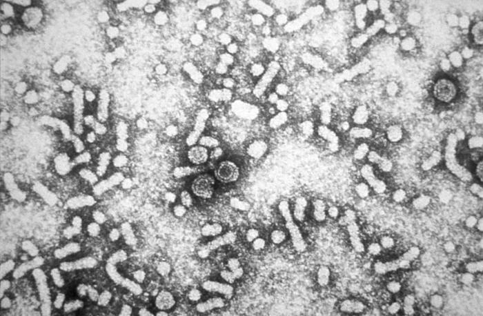
Hepatitis D virus(HDV) is the causative organism for Hepatitis D infection. HDV is only found in people who carry the hepatitis B virus. HDV may make a recent (acute) hepatitis B infection or an existing long-term (chronic) hepatitis B liver disease worse. It can even cause symptoms in people who carry hepatitis B virus but who never had symptoms. Hepatitis D infects about 15 million people worldwide. It occurs in 5% of people who carry hepatitis B. Risk factors include:
- Abusing intravenous (IV) or injection drugs
- Being infected while pregnant (the mother can pass the virus to the baby)
- Carrying the hepatitis B virus
- Men having sexual intercourse with other men
- Receiving many blood transfusions
Virology
Genome structure and similarities to viroids
The HDV genome exists as a negative sense, single-stranded, closed circular RNA. Because of a nucleotide sequence that is 70% self-complementary, the HDV genome forms a partially double stranded RNA structure that is described as rod-like.[2] With a genome of approximately 1700 nucleotides, HDV is the smallest "virus" known to infect animals. It has been proposed that HDV may have originated from a class of plant viruses called viroids.[3] Evidence in support of this hypothesis stems from the fact that both HDV and viroids exist as single-stranded, closed circular RNAs that have rod-like structures. Likewise, both HDV and viroids contain RNA sequences that can assume catalytically active structures called ribozymes. During viral replication, these catalytic RNAs are required in order to produce unit length copies of the genome from longer RNA concatamers. Finally, neither HDV nor viroids encode their own polymerase. Instead, replication of HDV and viroids requires a host polymerase that can utilize RNA as a template.[4] RNA polymerase II has been implicated as the polymerase responsible for the replication of HDV.[5][6] Normally RNA polymerase II utilizes DNA as a template and produces mRNA. Consequently, if HDV indeed utilizes RNA polymerase II during replication, it would be the only known pathogen capable of using a DNA-dependent polymerase as an RNA-dependent polymerase.
Life Cycle
To replicate efficiently, a virus requires the cooperation of the host cell at all stages of the replicative cycle:[7]
- Attachment
- Penetration
- Uncoating
- Provision of appropriate metabolic conditions for the synthesis of viral macromolecules
- Final assembly of viral subunits
- Release of new virions
- HDV also requires the presence of a helper hepadnavirus to provide the protein components for its own envelope. How HDV enters hepatocytes is still not known, but it may involve the interaction between HBsAg-L and a cellular receptor. The incoming HDV RNA is then transported into the nucleus, probably by the small form of delta antigen, HDAg-S. Binding of HDAg to RNA also protects the HDV RNAs from degradation.[7]
- HDV RNA replication is carried out by cellular RNA polymerase II, without a DNA intermediate, and without the help of HBV.
- RNA transcription is regulated:[7]
- Initially - mRNA(s) is(are) transcribed from the incoming minus-strand genome
- Later - after the translation of the mRNA to make essential replication proteins, there is a switch in the mode of RNA-directed RNA synthesis to facilitate replication of the RNA genome
- Three forms of RNA are made:
- Circular genomic RNA
- Circular complementary antigenomic RNA
- Linear polyadenylated antigenomic RNA - mRNA containing the open reading frame for the HDAg.
Synthesis of antigenomic RNA occurs in the nucleous, mediated by RNA polymerase I, whereas synthesis of genomic RNA takes place in the nucleoplasm, mediated by RNA Pol II.[8]
- Small (p24) form of HDAg (HDAg-S) - transactivator of HDV RNA replication
- Large (p27) form of HDAg (HDAg-L) - inhibits RNA synthesis and initiates virion assembly with HBsAg.
- HDV particles include:
These elements are only assembled in the presence of the hepatitis B virus (helper virus). HBsAg and HDAg-L are necessary and sufficient for virus assembly, whereas HDV RNA or HDAg-S are not required, but are present, in viral particles. The primary initiation event for HDV assembly is the interaction of HDAg-L with HBsAg. HDAg is localized in the nuclei while and HBsAg is present in the cytoplasm of the infected cells. The mechanism of interaction between these two proteins remains unknown. That are different models that try to explain the mechanisms of viral RNA transcription and replication.[7][12][13]
The Delta Antigens
A significant difference between viroids and HDV is that, while viroids produce no proteins, HDV produces two proteins called the small and large delta antigens (HDAg-S and HDAg-L, respectively). These two proteins are produced from a single open reading frame. They are identical for 195 amino acids and differ only by the presence of an additional 19 amino acids at the C-terminus of HDAg-L. Despite having 90% identical sequences, these two proteins play diverging roles during the course of an infection. HDAg-S is produced in the early stages of an infection and is required for viral replication. HDAg-L, in contrast, is produced during the later stages of an infection, acts as an inhibitor of viral replication, and is required for assembly of viral particles.
Evolution
Three genotypes (I-III) were originally described. Genotype I has been isolated in Europe, North America, Africa and some Asia. Genotype II has been found in Japan, Taiwan, and Yakutia (Russia). Genotype III has been found exclusively in South America (Peru, Colombia, and Venezuela). Some genomes from Taiwan and the Okinawa islands have been difficult to type but have been placed in genotype 2. However it is not known that there are at least 8 genotypes of this virus (HDV-1 to HDV-8).[14]Phylogenetic studies suggest an African origin for this pathogen.[15]
References
- ↑ "http://www.who.int/en/". External link in
|title=(help) - ↑ Saldanha JA, Thomas HC, Monjardino JP (1990). "Cloning and sequencing of RNA of hepatitis delta virus isolated from human serum". J. Gen. Virol. 71 ( Pt 7): 1603–6. doi:10.1099/0022-1317-71-7-1603. PMID 2374010. Unknown parameter
|month=ignored (help) - ↑ Elena SF, Dopazo J, Flores R, Diener TO, Moya A (1991). "Phylogeny of viroids, viroidlike satellite RNAs, and the viroidlike domain of hepatitis delta virus RNA". Proc. Natl. Acad. Sci. U.S.A. 88 (13): 5631–4. doi:10.1073/pnas.88.13.5631. PMC 51931. PMID 1712103. Unknown parameter
|month=ignored (help) - ↑ Taylor JM (2003). "Replication of human hepatitis delta virus: recent developments". Trends Microbiol. 11 (4): 185–90. doi:10.1016/S0966-842X(03)00045-3. PMID 12706997. Unknown parameter
|month=ignored (help) - ↑ Lehmann E, Brueckner F, Cramer P (2007). "Molecular basis of RNA-dependent RNA polymerase II activity". Nature. 450 (7168): 445–9. doi:10.1038/nature06290. PMID 18004386. Unknown parameter
|month=ignored (help) - ↑ Filipovska J, Konarska MM (2000). "Specific HDV RNA-templated transcription by pol II in vitro". RNA. 6 (1): 41–54. doi:10.1017/S1355838200991167. PMC 1369892. PMID 10668797. Unknown parameter
|month=ignored (help) - ↑ 7.0 7.1 7.2 7.3 7.4 "Hepatitis D" (PDF).
- ↑ Li, YJ (2006 Jul). "RNA-Templated Replication of Hepatitis Delta Virus: Genomic and Antigenomic RNAs Associate with Different Nuclear Bodies". Journal of virology. 80 (13): 6478–86. doi:10.1128/JVI.02650-05. PMC 1488965. PMID 16775335. Unknown parameter
|coauthors=ignored (help); Check date values in:|date=(help) - ↑ Dingle K, Bichko V, Zuccola H, Hogle J, Taylor J (1998). "Initiation of hepatitis delta virus genome replication". J Virol. 72 (6): 4783–8. PMC 110015. PMID 9573243.
- ↑ Lai MM (1995). "The molecular biology of hepatitis delta virus". Annu Rev Biochem. 64: 259–86. doi:10.1146/annurev.bi.64.070195.001355. PMID 7574482.
- ↑ Ryu WS, Bayer M, Taylor J (1992). "Assembly of hepatitis delta virus particles". J Virol. 66 (4): 2310–5. PMC 289026. PMID 1548764.
- ↑ Modahl LE, Lai MM (1998). "Transcription of hepatitis delta antigen mRNA continues throughout hepatitis delta virus (HDV) replication: a new model of HDV RNA transcription and replication". J Virol. 72 (7): 5449–56. PMC 110180. PMID 9621000.
- ↑ Fields, Bernard (2013). Fields virology. Philadelphia: Wolters Kluwer Health/Lippincott Williams & Wilkins. ISBN 1451105630.
- ↑ Celik I, Karataylı E, Cevik E; et al. (2011). "Complete genome sequences and phylogenetic analysis of hepatitis delta viruses isolated from nine Turkish patients". Arch. Virol. 156 (12): 2215–20. doi:10.1007/s00705-011-1120-y. PMID 21984217. Unknown parameter
|month=ignored (help) - ↑ Radjef N, Gordien E, Ivaniushina V, Gault E, Anaïs P, Drugan T, Trinchet JC, Roulot D, Tamby M, Milinkovitch MC, Dény P (2004) Molecular phylogenetic analyses indicate a wide and ancient radiation of African hepatitis delta virus, suggesting a deltavirus genus of at least seven major clades. J Virol 78(5):2537-2544