Hashimoto's thyroiditis chest x ray: Difference between revisions
| Line 15: | Line 15: | ||
Image:Pleural effusion.jpg|'''Pleural effusion''' Chest x-ray of a pleural effusion. The arrow A shows fluid layering in the right pleural cavity. The B arrow shows the normal width of the lung in the cavity - Case courtesy of Dr Vivek Pai, <a href="https://radiopaedia.org/">Radiopaedia.org</a>. From the case <a href="https://radiopaedia.org/cases/27112">rID: 27112</a> | Image:Pleural effusion.jpg|'''Pleural effusion''' Chest x-ray of a pleural effusion. The arrow A shows fluid layering in the right pleural cavity. The B arrow shows the normal width of the lung in the cavity - Case courtesy of Dr Vivek Pai, <a href="https://radiopaedia.org/">Radiopaedia.org</a>. From the case <a href="https://radiopaedia.org/cases/27112">rID: 27112</a> | ||
Image:Right side pleural effusion 2.png|Right side pleural effusion. A homogenous opacification is noted in the right lower zone. The right costophrenic angle is obliterated with a meniscus noted. - Source: https://www.cdc.gov/ | Image:Right side pleural effusion 2.png|Right side pleural effusion. A homogenous opacification is noted in the right lower zone. The right costophrenic angle is obliterated with a meniscus noted. - Source: https://www.cdc.gov/ | ||
File:Mohsin gif.gif|left|300px|thumb|'''Cardiomegaly''';Chest x-ray. Image courtesy of C. Michael Gibson MS. MD | |||
</gallery> | </gallery> | ||
Revision as of 17:04, 25 September 2017
|
Hashimoto's thyroiditis Microchapters |
|
Diagnosis |
|---|
|
Treatment |
|
Case Studies |
|
Hashimoto's thyroiditis chest x ray On the Web |
|
American Roentgen Ray Society Images of Hashimoto's thyroiditis chest x ray |
|
Risk calculators and risk factors for Hashimoto's thyroiditis chest x ray |
Editor-In-Chief: C. Michael Gibson, M.S., M.D. [1] Associate Editor(s)-in-Chief: Furqan M M. M.B.B.S[2]
Overview
The findings associated with Chest X ray in Hashimoto's thyroiditis are pleural effusion and cardiomegaly.
Chest X Ray
Chest X Ray in the patients with Hashimoto's thyroiditis may show:[1]
Gallery
-
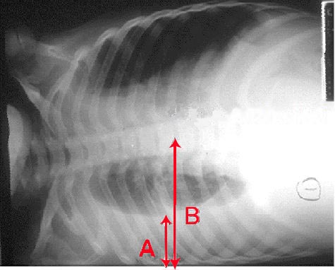
Pleural effusion Chest x-ray of a pleural effusion. The arrow A shows fluid layering in the right pleural cavity. The B arrow shows the normal width of the lung in the cavity - Case courtesy of Dr Vivek Pai, <a href="https://radiopaedia.org/">Radiopaedia.org</a>. From the case <a href="https://radiopaedia.org/cases/27112">rID: 27112</a>
-

Right side pleural effusion. A homogenous opacification is noted in the right lower zone. The right costophrenic angle is obliterated with a meniscus noted. - Source: https://www.cdc.gov/
-
Cardiomegaly;Chest x-ray. Image courtesy of C. Michael Gibson MS. MD


</gallery>
References
- ↑ Caturegli P, De Remigis A, Rose NR (2014). "Hashimoto thyroiditis: clinical and diagnostic criteria". Autoimmun Rev. 13 (4–5): 391–7. doi:10.1016/j.autrev.2014.01.007. PMID 24434360.