Editor-In-Chief: C. Michael Gibson, M.S., M.D. [1] Associate Editor(s)-in-Chief: Furqan M M. M.B.B.S [2]
Overview The histological analysis in Hashimoto's thyroiditis may show inflammatory cell infiltration and Hurthle cells. Fine needle aspiration cytology helps to differentiate between the benign and malignant nodules .
Other Diagnostic Studies The other diagnostic studies helpful in the diagnosis of Hashimoto's thyroiditis include histopathological analysis and fine needle aspiration cytology of thyroid gland.[1] [2]
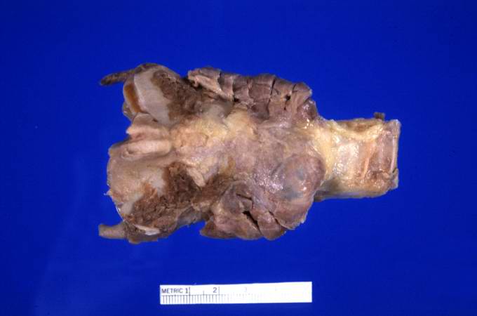
Gross Pathology The gland is usually diffusely enlarged, firm, and slightly lobular . The capsule is intact, and the cut surface is light-tan and has a slight lobular pattern.
At autopsy, significant subarachnoid hemorrhage from the ruptured berry aneurysm was documented. In addition, the thyroid gland was mildly enlarged and firm. On cut section, the tissue was slightly pale.
A gross photograph of thyroid gland taken at autopsy. The gland is only slightly enlarged and has a firm texture.Images courtesy of Professor Peter Anderson DVM Ph.D. and published with permission © PEIR, University of Alabama at Birmingham, Department of Pathology
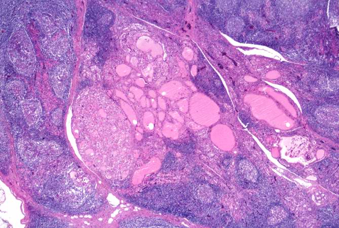
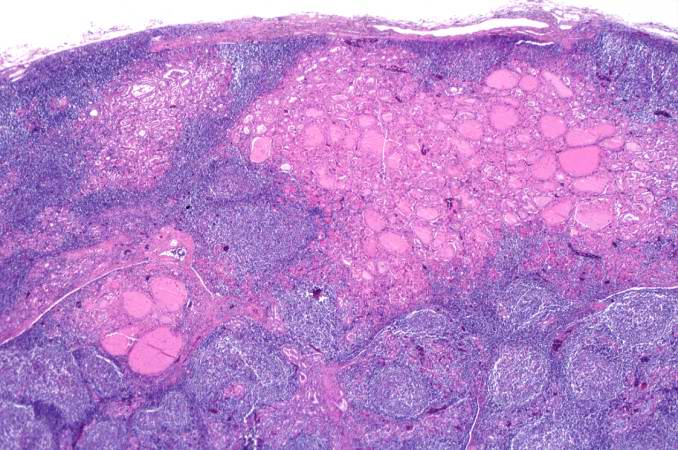
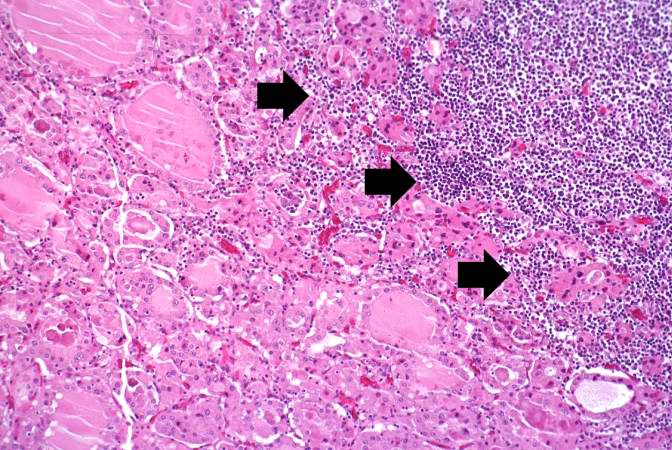
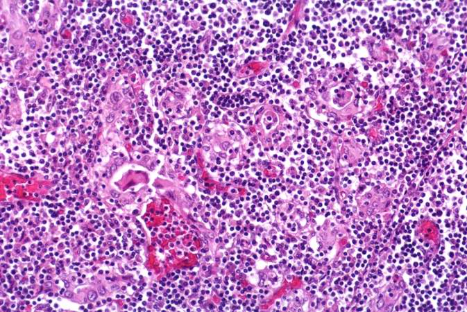
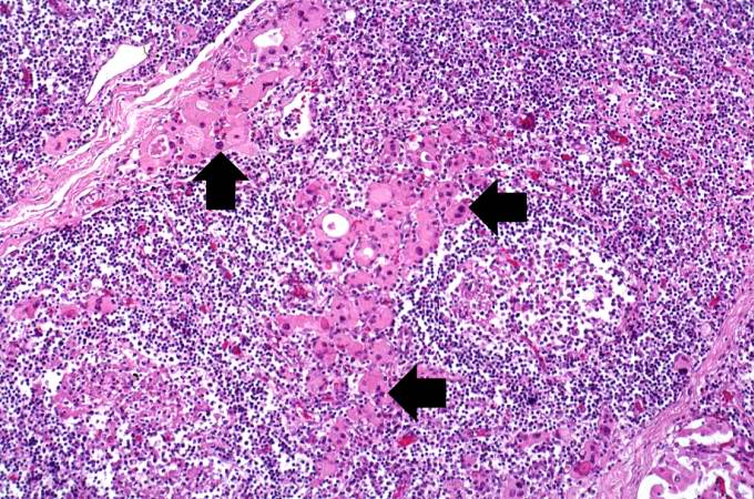
Microscopic Pathology Microscopically there is massive infiltration of the thyroid gland by lymphocytes and plasma cells . Germinal centers can often be seen in the gland . Thyroid follicles are usually absent and the few remaining follicles are devoid of colloid .
Photomicrograph shows germinal centers ; Case courtesy of Dr Andrew Ryan, Radiopaedia.org, rID: 17084
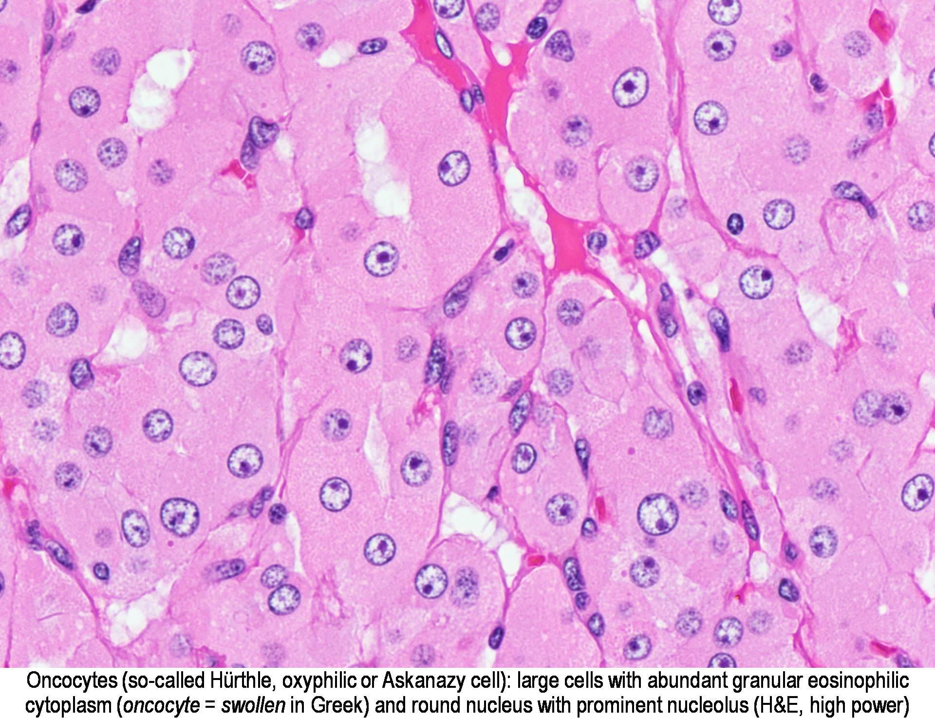
Micrograph of Hurthle cells Courtesy of PathologyOutlines.com; Source:[3]
This is a low-power photomicrograph of thyroid from this case. Note that the tissue is more cellular than one would expect and there does not appear to be normal colloid-filled blue spaces in this gland.Images courtesy of Professor Peter Anderson DVM Ph.D. and published with permission © PEIR, University of Alabama at Birmingham, Department of Pathology
This is a higher-power photomicrograph of thyroid from this case. Note a large number of blue-staining inflammatory cells in this tissue. These cells appear to be forming germinal centers. Some residual thyroid gland tissue can be seen in this section (arrows).Images courtesy of Professor Peter Anderson DVM Ph.D. and published with permission © PEIR, University of Alabama at Birmingham, Department of Pathology
This is another view of thyroid gland filled with inflammatory cells forming germinal centers (arrows).Images courtesy of Professor Peter Anderson DVM Ph.D. and published with permission © PEIR, University of Alabama at Birmingham, Department of Pathology
This is a higher-power photomicrograph of thyroid from this case showing the inflammatory cells and the residual thyroid tissue.Images courtesy of Professor Peter Anderson DVM Ph.D. and published with permission © PEIR, University of Alabama at Birmingham, Department of Pathology
This is another higher-power photomicrograph of thyroid from this case showing the inflammatory cells and the residual thyroid tissue.Images courtesy of Professor Peter Anderson DVM Ph.D. and published with permission © PEIR, University of Alabama at Birmingham, Department of Pathology
This is a high-power photomicrograph showing the inflammatory cells infiltrating into the residual thyroid tissue (arrows).Images courtesy of Professor Peter Anderson DVM Ph.D. and published with permission © PEIR, University of Alabama at Birmingham, Department of Pathology
This is a high-power photomicrograph showing the lymphocytes and plasma cells surrounding the thyroid gland epithelium.Images courtesy of Professor Peter Anderson DVM Ph.D. and published with permission © PEIR, University of Alabama at Birmingham, Department of Pathology
This high-power photomicrograph shows more clearly the lymphocytes and plasma cells surrounding the thyroid gland epithelium. Large, eosinophilic, degenerating thyroid gland cells (Hurthle cells) can be seen in this section (arrows).Images courtesy of Professor Peter Anderson DVM Ph.D. and published with permission © PEIR, University of Alabama at Birmingham, Department of Pathology
Fine needle aspiration cytology Fine needle aspiration is usually done under ultrasound guidance and the sample is sent for cytology . It helps to differentiate benign thyroid nodules from the malignant lesions.
References Template:WH
Template:WS
